
![]()
Ultrasound Case 049
Presentation
A 44 year old woman presents with right upper quadrant (RUQ) pain radiating through to her back. She has a fever and is tender on abdominal palpation. She has had several episodes of postprandial RUQ discomfort previously. You wonder if she has gallstones.
View 2
View 3
View 4

Describe and interpret these scans
IMAGE INTERPRETATION
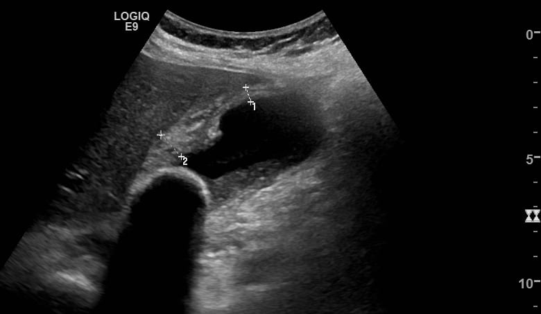
Image 1: Transverse view of the gallbladder fanning from the neck through to the fundus.
There is a large gallstone obstructing the neck of the gallbladder. It has a typical appearance with an echogenic anterior surface and dense dark posterior acoustic shadowing. The gallbladder wall is markedly thickened at 12mm (≤3mm) with striations reflecting oedema within the wall. There is some dependent sludge distal to the stone.
Image 2: Longitudinal view of the gallbladder with wall measurements.
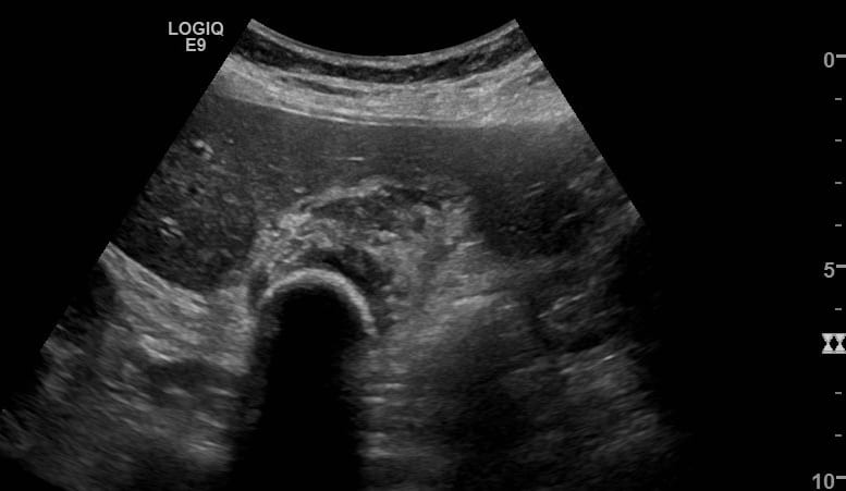
Image 3: Transverse image of gallbladder through the stone.
Image 4: Normal common bile duct.
CLINICAL CORRELATION
Acute cholecystitis
Biliary ultrasound is frequently complex with highly varied appearances and pathology. Accurate imaging and interpretation requires considerable expertise and experience.
The typical sonographic features of cholecystitis can include:
- Dilation of the gallbladder (transverse diameter >4cm)
- Gallstones and sludge particularly a stone lodged within the GB neck or cystic duct.
- Increased GB wall thickness (>3mm)
- Hyperaemia of the GB wall.
- Pericholecystic fluid
- Tenderness to transducer pressure over the gallbladder (sonographic Murphy’s sign)
Among the numerous pitfalls are:
- Incorrectly identifying the gallbladder – confusing it for bowel or vice versa.
- Missing a stone caught in the GB neck or cystic duct
- Attributing a thickened gallbladder wall to cholecystitis when it is due to another cause – and there are many for example adjacent inflammatory conditions such as hepatitis, pancreatitis or pyelonephritis.
- Not realizing the case is one of the many exceptions to the rules – such as acalculous cholecystitis – with no stones, or severe cholecystitis with absence of gallbladder wall hyperaemia, or a non tender gallbladder wall which occasionally happens in cholecystitis particularly in the elderly, or ascites thought to be inflammatory pericholecystic fluid…
[cite]
TOP 100 ULTRASOUND CASES

An Emergency physician based in Perth, Western Australia. Professionally my passion lies in integrating advanced diagnostic and procedural ultrasound into clinical assessment and management of the undifferentiated patient. Sharing hard fought knowledge with innovative educational techniques to ensure knowledge translation and dissemination is my goal. Family, wild coastlines, native forests, and tinkering in the shed fills the rest of my contented time. | SonoCPD | Ultrasound library | Top 100 | @thesonocave |
