![]()
Ultrasound Case 058
Presentation
A 71 year old man presents with severe back pain and hypotension. You suspect AAA.
View 2
Describe and interpret these scans
IMAGE INTERPRETATION
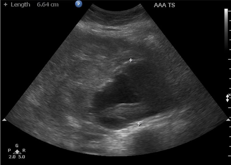
Image 1: Transverse view of the epigstrium sliding from xiphisternum to umbilicus. As the scan moves inferiorly the typical appearance of an abdominal aortic aneurysm with intraluminal thrombus becomes apparent. Outside the aorta is a large retroperitoneal haematoma.
Image 2: Transverse view just above umbilicus.

CLINICAL CORRELATION
Ruptured abdominal aortic aneurysm with retroperitoneal haematoma
Retroperitoneal haematoma may be anterior to the aorta, to the left (usually) or the right. This patient had had pain for several hours and the large retroperitoneal haematoma has the characteristic heterogeneous appearance – bits of black, bits of grey, bits of white.
[cite]
TOP 100 ULTRASOUND CASES

An Emergency physician based in Perth, Western Australia. Professionally my passion lies in integrating advanced diagnostic and procedural ultrasound into clinical assessment and management of the undifferentiated patient. Sharing hard fought knowledge with innovative educational techniques to ensure knowledge translation and dissemination is my goal. Family, wild coastlines, native forests, and tinkering in the shed fills the rest of my contented time. | SonoCPD | Ultrasound library | Top 100 | @thesonocave |