![]()
Ultrasound Case 071
Presentation
A 24 year old woman presents with generalised abdominal discomfort. She is concerned she looks pregnant but numerous pregnancy tests have been negative.
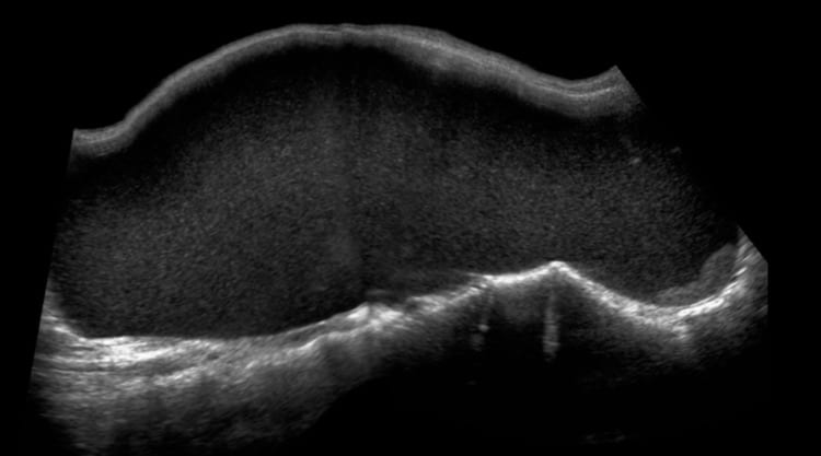
View 2: Panoramic view longitudinal mid abdomen
Describe and interpret these scans
IMAGE INTERPRETATION
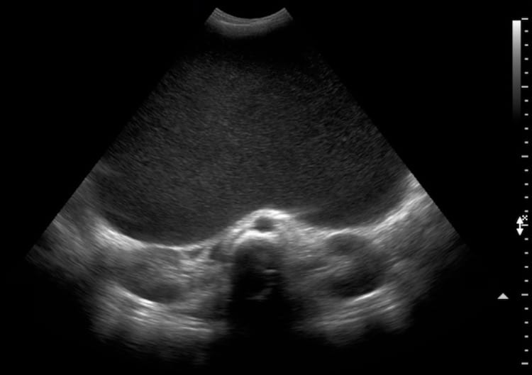
Image 1: Transverse view with the transducer midway between xiphisternum and umbilicus.
Posteriorly the aorta, IVC, vertebral body and body kidneys are seen. Filling most of the abdomen and displacing bowel into the upper abdomen is a large cystic structure filled with homogeneous low-level echoes sometimes described as a “ground-glass” appearance. A cyst this large giving this appearance is almost always ovarian in origin, and a mucinous cystadenoma the cause in this case.

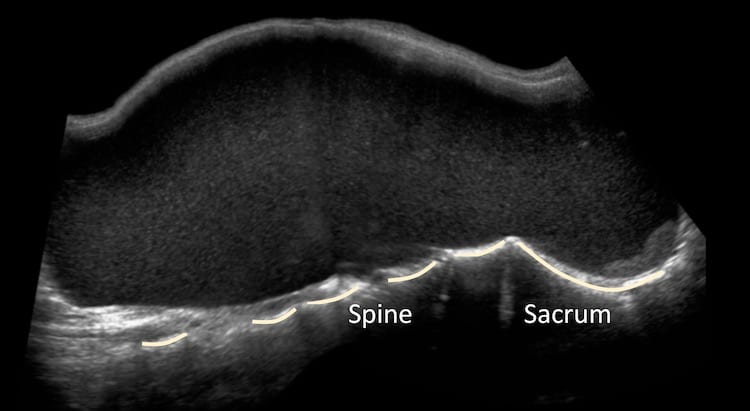
Image 2: Panoramic view of the abdomen in longitudinal section.
The large cyst is again demonstrated filling the pelvis and abdomen. Posteriorly the sacrum and vertebral bodies are seen.
CLINICAL CORRELATION
Mucinous cystadenoma
The mucinous cystadenoma is an ovarian epithelial tumour. Although often large it is benign and has excellent prognosis.
Mucin containing tumours range from the benign mucinous cystadenoma through borderline mucinous tumors to malignant mucinous cystadenocarcinoma of the ovary.
The appearance of homogeneous fine low-level echoes is not infrequently encountered in ultrasound and can be caused by mucin, haemorrhage, pus, or highly proteinaceous fluid.
[cite]
TOP 100 ULTRASOUND CASES

An Emergency physician based in Perth, Western Australia. Professionally my passion lies in integrating advanced diagnostic and procedural ultrasound into clinical assessment and management of the undifferentiated patient. Sharing hard fought knowledge with innovative educational techniques to ensure knowledge translation and dissemination is my goal. Family, wild coastlines, native forests, and tinkering in the shed fills the rest of my contented time. | SonoCPD | Ultrasound library | Top 100 | @thesonocave |