
![]()
Ultrasound Case 093
Presentation
A 9 year old boy presented with fever and right flank pain. The concern was pyelonephritis. Look at the first image and see what you think.
View 2: RUQ view longitudinal
View 3: RUQ view transverse
View 4: RUQ view transverse
Describe and interpret these scans
IMAGE INTERPRETATION
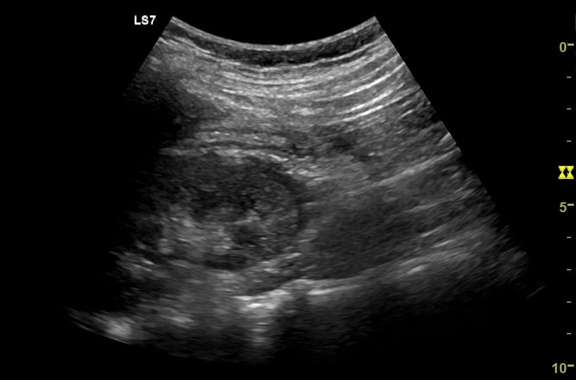
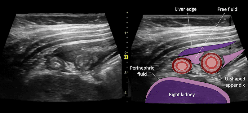
Image 1: Longitudinal image lower pole of the right kidney. This shows a rim of perinephric fluid, initially raising suspicion for pyelonephritis.

Image 2: Video longitudinal RUQ through Morrison’s pouch.
Image 3 & 4: Video in transverse through RUQ.
An inflamed loop of bowel sits in Morrison’s pouch. Closer inspection demonstrates it has typical bowel wall signature, is U-shaped and has a blind ending tip. It is surrounded by echogenic inflamed mesenteric fat and there is a small amount of associated free fluid. A single small round hypoechoic mesenteric lymph node is also seen. A reactive rim of perinephric fluid is evident.
Appendicitis with the appendix lying in Morrison’s pouch causing surrounding inflammatory change.
CLINICAL CORRELATION
Appendicitis – with the appendix lying in Morrison’s pouch
The initial image focused on the right kidney and demonstrated a rim of perinephric fluid. The clinical suspicion was of pyelonephritis.
Ultrasound is relatively poor at demonstrating uncomplicated pyelonephritis as there is generally no gross anatomical change. Findings can include echogenic debris in the collecting system, areas of increased or decreased parenchymal echogenicity, perinephric fluid or reduced perfusion demonstrated on Colour Doppler. The primary role of ultrasound is to assess for causes and complications. Ultrasound can exclude underlying anatomical abnormality, ureteric obstruction, renal abscess formation, or perinephric collections.
The possibility of an early perinephric collection was raised in this case, however changing one’s focus to view the abnormal inflamed bowel adjacent to the kidney gave the answer.
The bowel changes are typical of appendicitis. There is a tubular structure with bowel wall signature and blind ending tip with surrounding echogenic mesenteric fat and a small amount of free fluid. It measures up to 10mm across and peristalsis is not seen.
A small rim of perinephric fluid is similar to “stranding” seen on CT scanning. In the Emergency setting it is usually seen in the settings of renal inflammation or acute obstruction. In this case the adjacent inflammatory change of appendicitis was enough to cause the rim of perinephric fluid.
[cite]
TOP 100 ULTRASOUND CASES

An Emergency physician based in Perth, Western Australia. Professionally my passion lies in integrating advanced diagnostic and procedural ultrasound into clinical assessment and management of the undifferentiated patient. Sharing hard fought knowledge with innovative educational techniques to ensure knowledge translation and dissemination is my goal. Family, wild coastlines, native forests, and tinkering in the shed fills the rest of my contented time. | SonoCPD | Ultrasound library | Top 100 | @thesonocave |