![]()
Ultrasound Case 095
Presentation
A 44 year old woman presents with calf pain after a long hike followed by a long flight. Her upper medial calf is particularly tender. You consider Baker’s cyst; calf muscle tear; and DVT the most likely differentials.
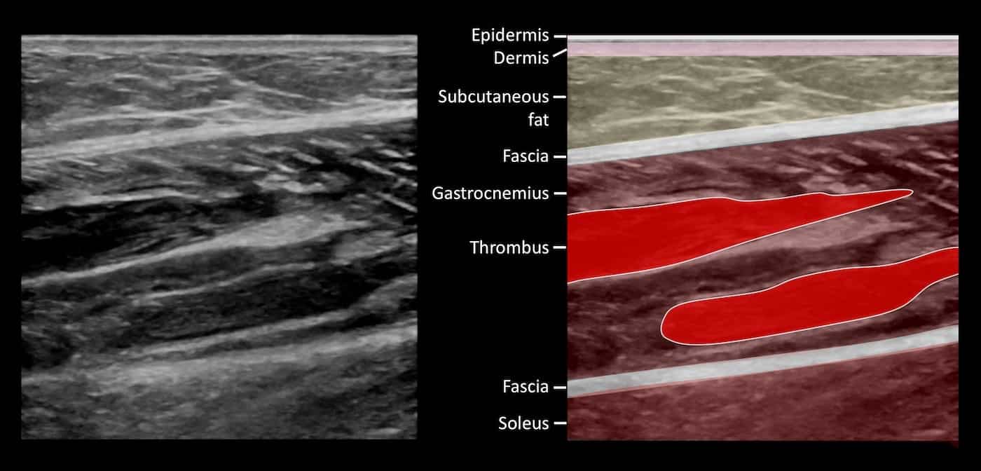
Image 2: Longitudinal image of medial gastrocnemius muscle
Describe and interpret these scans
IMAGE INTERPRETATION
Image 1: Transverse view of left medial gastrocnemius muscle running the probe from proximal to distal.
The normally compressible and small gastrocnemius veins are dilated and filled with echogenic thrombus. In this patient the gastrocnemius muscle vein is paired and both contain thrombosis. The thrombus is extensive but does not reach the posterior tibial or popliteal veins. A thin rim of hypoechoic fluid – oedema is seen around the gastrocnemius muscle.
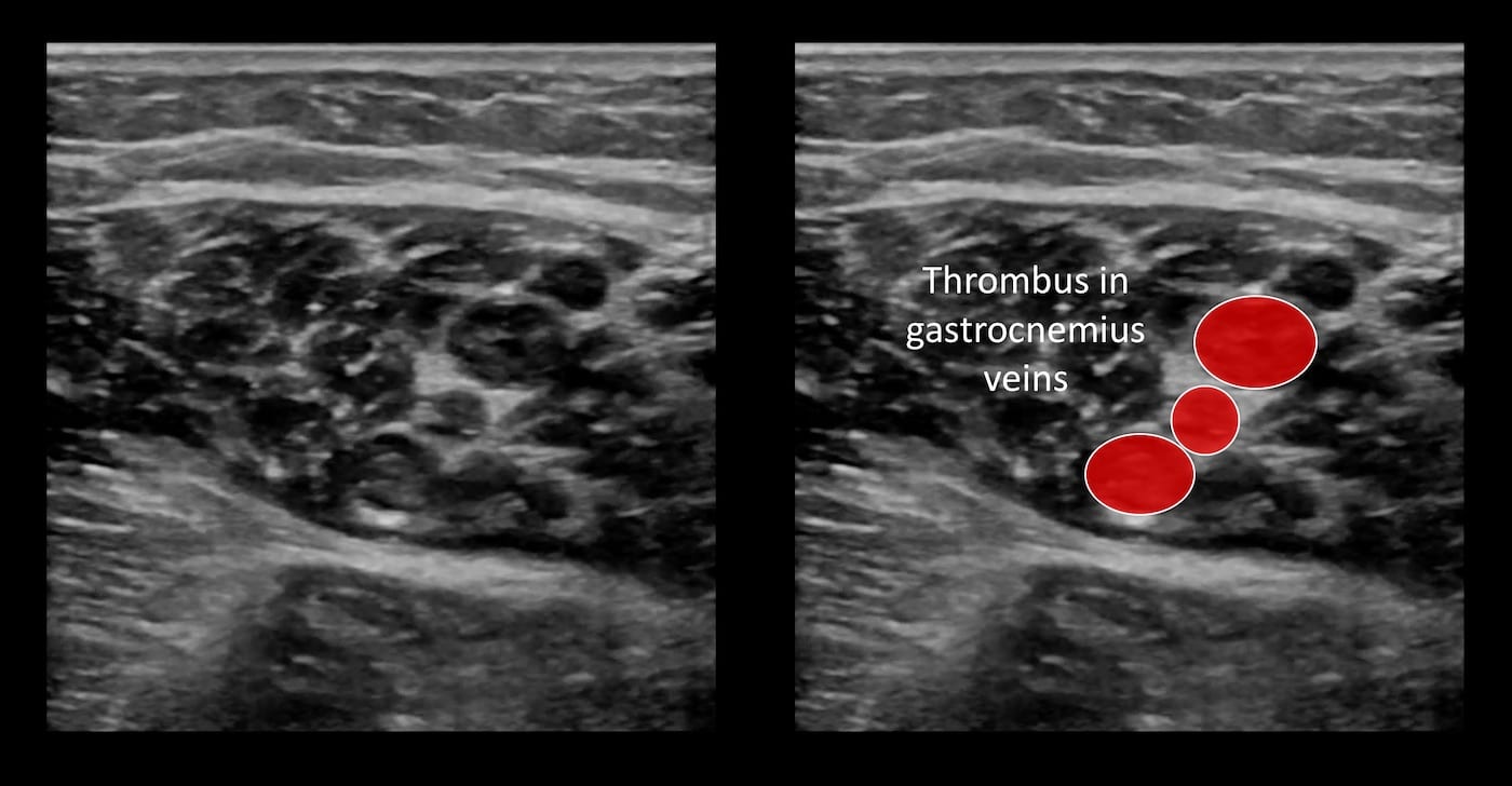
Image 2: Longitudinal image of medial gastrocnemius muscle demonstrating vein thrombosis.
CLINICAL CORRELATION
Isolated calf vein muscle thrombosis
A full DVT scan includes examining veins below the knee.
Examine for echogenic thrombus within the vein. In the acute case the vein is often distended.
Assess for compressibility by slowly pressing with the transducer. A non-thrombosed vein will be readily compressible.
Assess for flow variation – respiratory variation and augmentation both alter flow within a vein and can give information about obstruction proximal and distal to the site of examination. This is demonstrated using colour and pulse wave Doppler.
Isolated thrombus in the medial head of gastrocnemius is common. In this case it was extremely tender and therapeutic anticoagulation was commenced with re-imaging at 6 weeks, which demonstrated complete resolution. The thrombus was provoked and anticoagulation ceased at 6 weeks with no further recurrence.
The optimal ultrasound examination, classification and management of DVT particularly isolated distal deep vein thrombosis and isolated calf muscle vein thrombosis remains controversial and are tailored according to local expertise, resources, clinical parameters, and patient preference.
[cite]
TOP 100 ULTRASOUND CASES

An Emergency physician based in Perth, Western Australia. Professionally my passion lies in integrating advanced diagnostic and procedural ultrasound into clinical assessment and management of the undifferentiated patient. Sharing hard fought knowledge with innovative educational techniques to ensure knowledge translation and dissemination is my goal. Family, wild coastlines, native forests, and tinkering in the shed fills the rest of my contented time. | SonoCPD | Ultrasound library | Top 100 | @thesonocave |