
![]()
Ultrasound Case 103 Fire and Brimstone
Presentation
A 38 year old woman presents with midcycle LIF pain. It is colicky and you are asked to scan her pelvis.

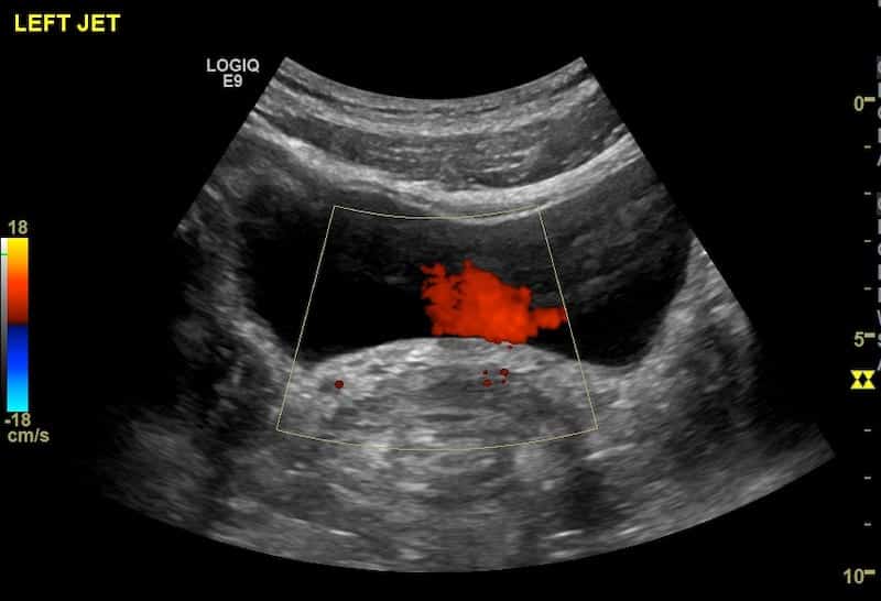
View 2: Transverse Bladder
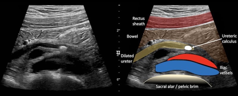
View 3: Left mid ureter
View 4: Left mid ureter with colour Doppler
Describe and interpret these scans
IMAGE INTERPRETATION
Image 1: Left kidney. Normal left kidney with no hydronephrosis.
Image 2: Transverse Bladder. Normal left ureteric jet; this excludes a completely obstructing ureteric calculus, but sometimes a stone does not cause complete ureteric obstruction.
Image 3: Left mid ureter. The ureter crosses the iliac vessels at the pelvic brim. A 5mm echogenic shadow casting lesion is seen within the lumen of a slightly distended ureter. The cause of her pain is ureteric colic.
Image 4: With colour Doppler the vessels are interrogated. Flow is demonstrated in the iliac vessels, not in the ureter, and the stone demonstrates “twinkle” artefact.
CLINICAL CORRELATION
Left ureteric colic
Renal stones generally cause symptoms when they pass into the ureter. They are known to get stuck at three points – the pelvi-ureteric junction (PUJ), the mid ureter at the pelvic brim as the ureter crosses the iliac vessels, and distally at the vesico-ureteric junction (VUJ).
In this case the stone is seen at the mid ureter.
Ultrasound technique: I always search for the mid ureter in patients with suspected renal colic. With the probe placed in the saggital plane I slide the probe along the iliac vessels from origin to groin. A dilated ureter will be seen as a hypoechoic tubular structure running obliquely over the iliac vessels at their mid point. A non dilated normal ureter is usually not seen.
TOP 100 ULTRASOUND CASES

An Emergency physician based in Perth, Western Australia. Professionally my passion lies in integrating advanced diagnostic and procedural ultrasound into clinical assessment and management of the undifferentiated patient. Sharing hard fought knowledge with innovative educational techniques to ensure knowledge translation and dissemination is my goal. Family, wild coastlines, native forests, and tinkering in the shed fills the rest of my contented time. | SonoCPD | Ultrasound library | Top 100 | @thesonocave |

Thank you so much for these amazing learning cases!! For those of us just learning POCUS, photos of probe placement would be so wonderfully helpful 🙂
VERY USEFULL SITE INSTRUCTIVE, SUPERB QUALITY PICTURES
This is the best website i have ever seen… Amazing..its improving my interpretation skills too much… Thank you so much