Ultrasound Case 112
Presentation
A 30 year old woman who is currently 30 weeks gestation presents to the ED with abrupt, severe right loin pain. She has moderate haematuria but is otherwise well. Her bloods are unremarkable.
She has had a normal pregnancy thus far and has no significant medical history.
A focused obstetric US shows a healthy 30/40 singleton pregnancy in cephalic presentation.
The kidneys were then evaluated using ultrasound


Questions
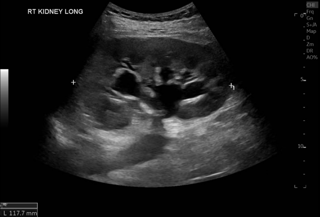
Interpret the LONG VIEW of the right kidney
The right kidney images demonstrate moderate hydronephrosis with clubbing of the calyces and dilation of the renal pelvis.
In pregnancy mild to moderate hydronephrosis can be normal and asymptomatic.
Up to 90% of women may have physiological hydronephrosis seen on ultrasound, more frequently in the second and third trimesters as the gravid uterus compresses the ureter at the pelvic brim.
The right kidney is more prone to gestational hydronephrosis than the left.

Interpret View 2: LONGITUDINAL VIEW of the INFERIOR POLE of the RIGHT KIDNEY
The inferior pole of the kidney is seen with anechoic fluid surrounding the kidney and extending into the retroperitoneal space anterior to the psoas muscle.
There is also a significant volume of anechoic perinephric fluid seen. This is not in keeping with physiological pregnancy-related hydronephrosis. This is likely to represent rupture of one of the calyces and small urinoma as a result. This is a relatively rare finding in cases of ureteric obstruction.

The patient underwent urgent MRI

Interpret the MRI
The patient underwent urgent MRI – this shows the same degree of hydronephrosis and perinephric fluid collection. No ureteric stone was identified.
Clinical Comment
It is thought that the ureteric obstruction was the result of the gravid uterus obstructing the ureter as it passes over the pelvic brim with subsequent increased intracalyceal pressure and minor rupture of the collecting system.
This is a rare complication of pregnancy with only 40 cases reported in the literature.
CONCLUSION:
Patient was booked for a JJ stent to relieve the obstruction.
REFERENCES
- Vega-Figueroa K, Silva-Melendez P, Figueroa-Gonzalez R, Colom-Diaz A, Gonzalez K. A Spontaneous Renal Calyceal Rupture Mimicking Physiologic Changes of Pregnancy and Other Common Pathologies. Cureus. 2023 Nov 18;15(11):e49006.
TOP 100 ULTRASOUND CASES
GP working in Broome in the NW of Western Australia. I work as a hospital DMO (District Medical Officer) doing Emergency, Anaesthestics, some Obstetrics and a lot of miscellaneous primary care | @broomedocs | BroomeDocs |
An Emergency physician based in Perth, Western Australia. Professionally my passion lies in integrating advanced diagnostic and procedural ultrasound into clinical assessment and management of the undifferentiated patient. Sharing hard fought knowledge with innovative educational techniques to ensure knowledge translation and dissemination is my goal. Family, wild coastlines, native forests, and tinkering in the shed fills the rest of my contented time. | SonoCPD | Ultrasound library | Top 100 | @thesonocave |


