![]()
A Woman of Singular Vision
aka Ophthalmology Befuddler 027
A 56 year-old female presents with sudden onset loss of vision in her right eye. She a past medical history of hypertension, hyperlipidema and medication-controlled diabetes mellitus type 2. Her medications include aspirin, ramipril, atorvastation and metformin. On examination she has 6/60 vision in her right eye.
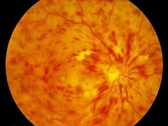
You perform fundoscopy and observe the following appearance:
Questions
Q1. What is the likely diagnosis?
Answer and interpretation
Central retinal vein occlusion (CRVO)
Sudden painless loss of vision, in a patient with risk factors and a ‘blood and thunder’ retinal appearance.
Q2. What are the predisposing factors and associated conditions?
Answer and interpretation
There are lots:
- glaucoma
- old age
- hypertension
- diabetes mellitus
- hypercoagulable state
- atherosclerosis (vein is compressed by adjacent artery)
- retrobular compressive lesions (e.g. thyroid disease, orbital tumour)
- vasculitis
Q3. What features on history and examination should be looked for?
Answer and interpretation
History:
- sudden and painless loss of vision
- assess for risk factors/ underlying causes (see Q2)
Examination:
- Visual acuity — variable depending on severity and duration since onset
- A Marcus-Gunn pupil may be present if ischemic CRVO (relative afferent pupillary defect = RAPD)
- Red reflex: may be abnormal
Fundoscopy: (Large areas of hemorrhage)
- non-ischemic CRVO: dilated tortuous veins, retinal hemorrhages, cotton wool spots, retinal edema, disc swelling.
- ischemic CRVO (more severe): classic ‘blood and thunder’ appearance from widespread hemorrhages that obscure most fundal details. Neovascularisation.
Q4. What is the management?
Answer and interpretation
- Refer to an ophthalmologist — photocoagulation may be performed if there is neovascularisation.
- Refer to a physician for ongoing work-up and treatment of underlying causes
- Screen for risk factors (cardiovascular disease, diabetes, vasculitis, etc)
- Consider low-dose aspirin (unproven)
Q5. How does branch retinal vein occlusion differ from this condition?
Answer and interpretation
A branch retinal vein occlusion only affects a sector of the retina corresponding to the distribution of the affected branch. Visual loss is limited to a segment of the visual field.
References
- Ehlers JP, Shah CP, Fenton GL, Hoskins EN. The Wills Eye Manual: Office and Emergency Room Diagnosis and Treatment of Eye Disease Lippincott Williams & Wilkins
- NSW Statewide Opthalmology Service. Eye Emergency Manual — An illustrated Guide. [Free PDF]
OPHTHALMOLOGY BEFUDDLER
Chris is an Intensivist and ECMO specialist at The Alfred ICU, where he is Deputy Director (Education). He is a Clinical Adjunct Associate Professor at Monash University, the Lead for the Clinician Educator Incubator programme, and a CICM First Part Examiner.
He is an internationally recognised Clinician Educator with a passion for helping clinicians learn and for improving the clinical performance of individuals and collectives. He was one of the founders of the FOAM movement (Free Open-Access Medical education) has been recognised for his contributions to education with awards from ANZICS, ANZAHPE, and ACEM.
His one great achievement is being the father of three amazing children.
On Bluesky, he is @precordialthump.bsky.social and on the site that Elon has screwed up, he is @precordialthump.
| INTENSIVE | RAGE | Resuscitology | SMACC