


Capener triangle
Capener triangle (Capener sign; triangular sign) is the normal dense triangular overlap of the inferomedial femoral neck/metaphysis with the posterior acetabular wall on an AP pelvic radiograph. The overlap is reduced or lost in slipped capital femoral epiphysis (SCFE).
The Capener triangle is an AP radiographic sign used in the assessment of slipped capital femoral epiphysis (SCFE). In the normal growing hip, part of the inferomedial femoral neck lies intra-articularly and is projected over the posterior wall of the acetabulum, producing a triangular area of increased radiographic density.
With SCFE, displacement between the capital epiphysis and femoral neck reduces this acetabular–metaphyseal overlap. The triangle therefore becomes smaller or disappears. The finding is best regarded as a supplementary sign rather than a stand-alone diagnostic one. Comparison with the opposite hip and appropriate lateral imaging remain important.
History
1956 – Norman Capener made an anatomical observation while discussing development and biomechanics of the hip. Capener did not describe a triangular density or name a radiographic sign.
Too often the capital epiphysis and the caput femorale are regarded as identical structures. The caput femorale, however, is partly diaphyseal in origin as may be seen clearly in infancy and adolescence. In adolescence recognition of this feature may help in the early diagnosis radiologically of epiphyseal coxa vara in which the diaphyseal portion of the caput may appear to be excluded from the acetabulum.
1960 – Frederick C. Durbin worked with Capener at the Princess Elizabeth Orthopaedic Hospital in Exeter. He expanded on Capener’s initial observation in his publication Treatment of slipped upper femoral epiphysis
It is not sufficiently realised that the caput femorale comprises more than the capital epiphysis. Capener (1956) has shown that the exclusion from the acetabulum of this articular part of the diaphysis … is amongst the earliest radiological signs of slipping
Durbin 1960
Durbin illustrated how the proximal femoral diaphysis normally extends medially beneath the capital epiphysis and how downward and posterior slipping excludes this articular component from the acetabulum. He attributed the observation to Capener and considered it one of the earliest radiographic signs of slipping.
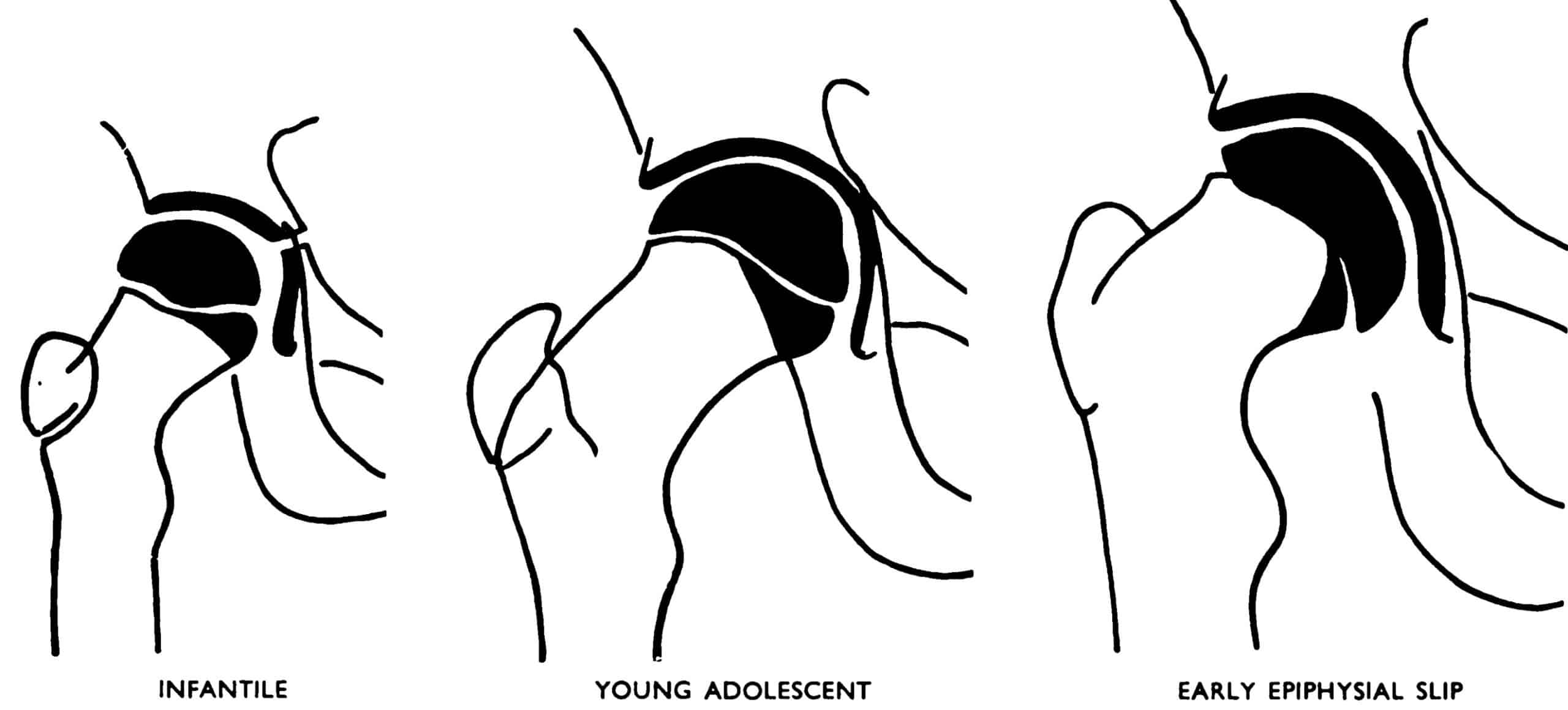
1962 – Philip Jacobs published A Note on the Diagnosis of Early Adolescent Coxa Vara (Slipped Epiphysis). He converted the anatomical observation into a more practical radiographic sign, which he termed “extrusion of the diaphyseal component of the caput femorale from the acetabular contour”.
His illustration depicts the normal diaphyseal component of the femoral head lying within the acetabulum as a white triangular area.
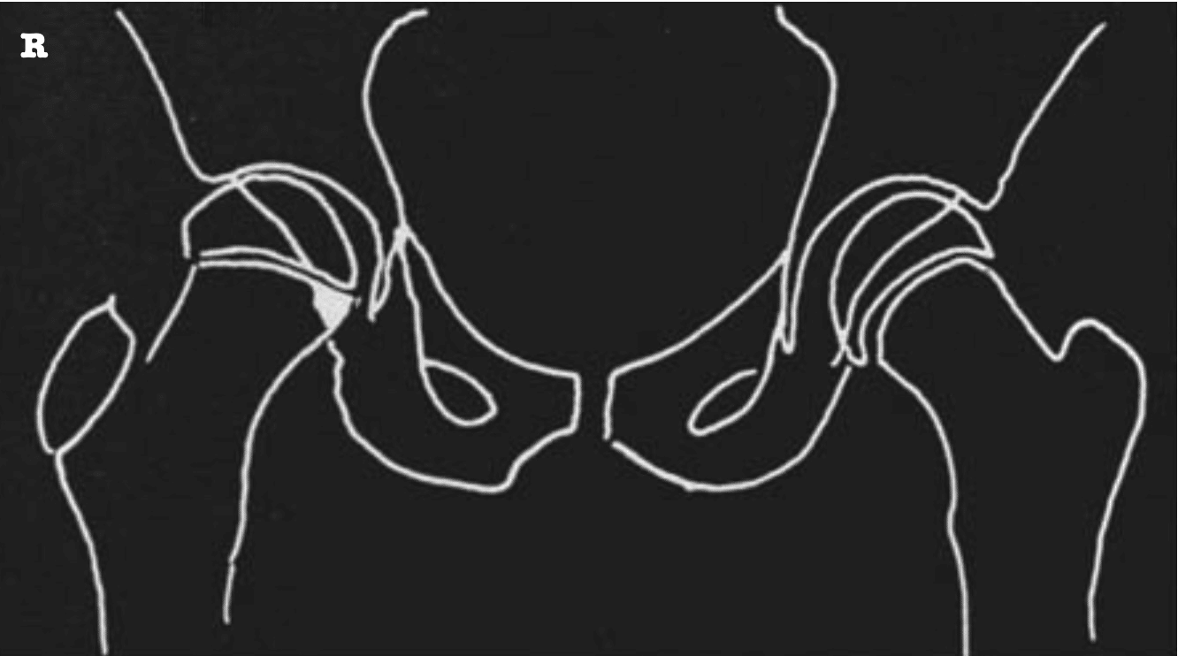
Jacobs reviewed 24 hips with early slipped epiphysis. The sign was present in all 24, and in four no other abnormality could be detected on the AP radiograph. He concluded that the sign was supplementary to other radiographic features and that a lateral view remained necessary to assess posterior slip.
With incomplete extrusion, comparison with the contralateral hip was required. Bilateral minimal slips could theoretically be misleading, while the uncommon upward slip associated with pre-existing coxa valga could invalidate the sign
1968 – Stewart M. Scham described a “dense triangle” normally produced by overlap of the lower acetabulum and the diaphyseal portion of the femoral neck. In SCFE this triangle was absent. He reported the finding was the only radiographic sign in two hips in his series of 44 hips.
1974 – Scham published the short paper The triangular sign in the early diagnosis of slipped capital femoral epiphysis. He described the normal inferomedial femoral neck as intra-articular and overlying the posterior acetabular wall, creating a dense triangular opacity. In most SCFE cases the relevant portion of the neck lies lateral to the acetabulum and the triangle is lost.
Scham did not claim the observation as his own and referenced Capener as first describing the sign in 1956 and discussed the subsequent work of Durbin and Jacobs.
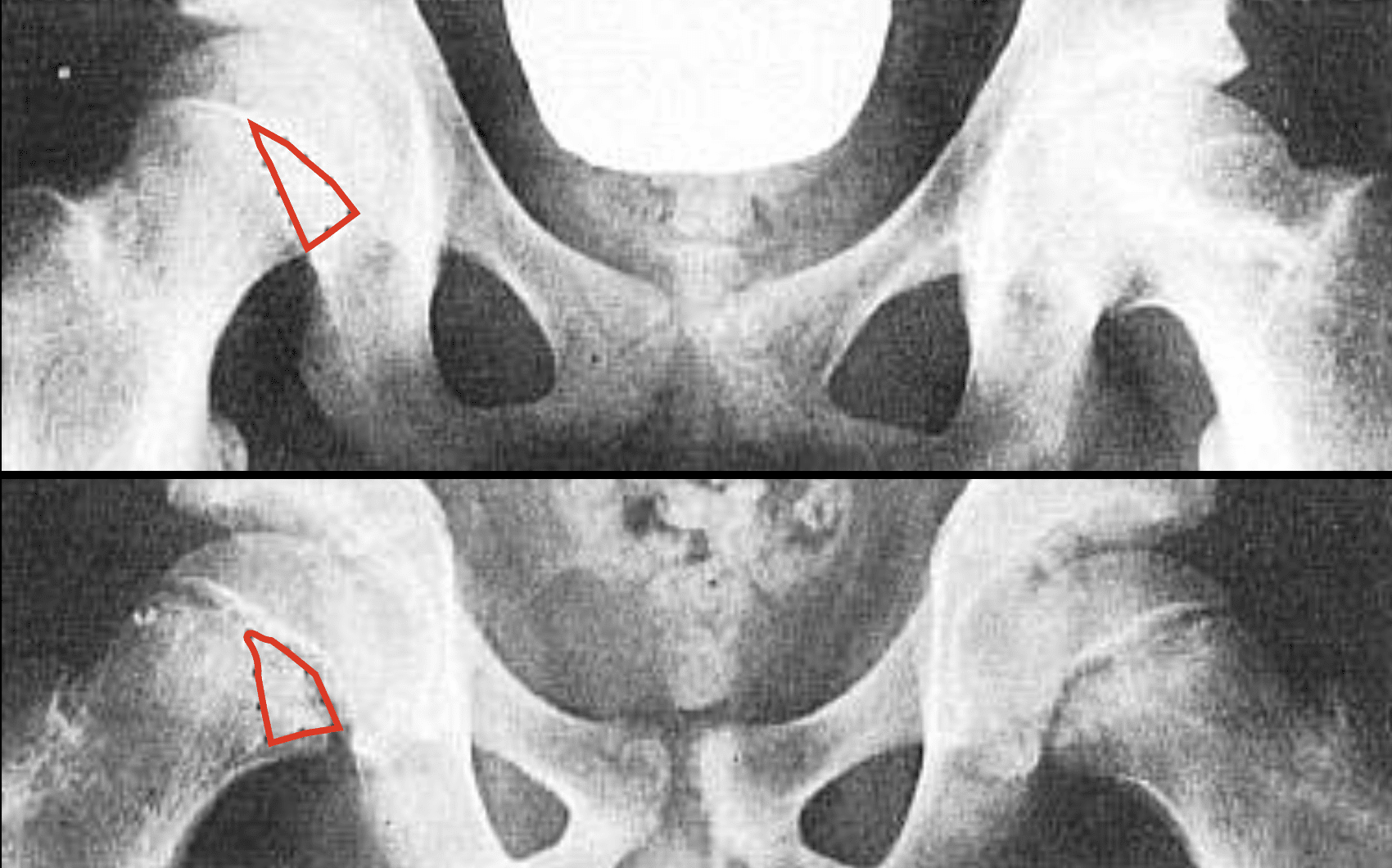
the triangle is outside the acetabulum
Eponymythology
Capener’s original description did not identify a “triangle”. In 1956 he described the developmental anatomy underlying the sign and noted exclusion of the diaphyseal component of the femoral head from the acetabulum in epiphyseal coxa vara.
Durbin provided a clarification and illustration of Capener’s paragraph in 1960. Jacobs depicted the resulting triangular overlap and evaluated its diagnostic value in 1962. Scham described the “dense triangle” in 1968 and the “triangular sign” in 1974.
Later use of Capener triangle and Capener sign attributes the underlying observation to Capener, rather than reproducing terminology introduced by Capener himself.
Some teaching texts present the Capener and Scham signs separately. Capener as loss of overlap between the posterior acetabular margin and medial metaphysis, and Scham as disappearance of the resulting triangular density. Historically, however, these are two descriptions of the same projectional phenomenon rather than independent signs.
Associated Persons
- Norman Leslie Capener (1898-1975)
Alternative names
- Capener sign
- triangular sign
- Scham sign — later attribution
- loss of Capener triangle
References
Historical references
- Capener NL. Reconstructive Surgery of the Hip Joint. In: Modern Trends in Orthopaedics (2nd series), edited by Sir Harry Platt, 1956
- Durbin FC. Treatment of slipped upper femoral epiphysis. J Bone Joint Surg Br. 1960;42-B(2):289-302
- Jacobs P. A Note on the Diagnosis of Early Aolescent Coxa Vara (Slipped Epiphysis), British Journal of Radiology, 1962; 35(417): 619–621
- Scham SM. Capital Femoral Epiphysiolysis Treated by Plugging with Cortical Beef Bone. Acta Orthopaedica Scandinavica, 1968; 39: 1-3, 171-181
- Scham SM. The triangular sign in the early diagnosis of slipped capital femoral epiphysis. Clin Orthop Relat Res. 1974;(103):16-7.
Eponymous term review
- Foster K. The limping child, Imaging, 2004; 16(2): 153–160
- How Kit N. Slipped capital femoral epiphysis (SCFE): what we should not miss on x-rays. ECR 2017
- Samelis PV, Loukas C, Kantanoleon S, Lalos H, Anoua N, Kolovos P, Georgiou F, Konstantinou AL. Causes of Delayed Diagnosis of Slipped Capital Femoral Epiphysis: The Importance of the Frog Lateral Pelvis Projection. Cureus. 2020 Apr 18;12(4):e7718.
- Kyle E, Cadogan M. Hip and pelvic lines and arcs. LITFL
eponymictionary
the names behind the name

Sujit Iyer, MD, is the National Director of Pediatric Services at US Acute Care Solutions and a Professor of Pediatrics and Pediatric EM Fellowship Director at UT Austin Dell Medical School. He completed his medical training and pediatrics residency at Baylor College of Medicine / Texas Children’s Hospital, followed by a Pediatric Emergency Medicine fellowship at Children’s Hospital of Philadelphia. Dr. Iyer focuses on emergency department operations, pediatric readiness in community hospitals, and curriculum development, lecturing nationally on quality improvement and pediatric emergency medicine best practices.

BA MA (Oxon) MBChB (Edin) FACEM FFSEM. Emergency physician, Sir Charles Gairdner Hospital. Passion for rugby, medical history, medical education, and asynchronous learning #FOAMed evangelist. Editor-in-Chief and Webmaster of Life in the Fast lane, LITFL | On Call 4e| Eponyms | Books | Horology |