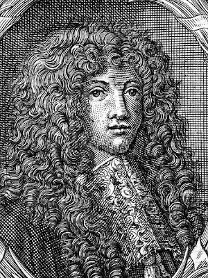
Caspar Bartholin Secundus
Biography
- Born 10 September 1655 in Copenhagen, Denmark
- 1674 – Professor of philosophy at the University of Copenhagen
- 1678 – Doctor of medicine (conferred upon him by his father Thomas Bartholin)
- 1691 – Supreme Court Assessor
- 1719 – Attorney general
- 1724 – Deputy for finance
- 1729 – The Order of the Dannebrog (Dannebrogordenen)
- Died 1 June 1738
Medical Eponyms
Bartholin glands (1677)
[aka Bartholin’s glands; greater vestibular glands; vulvovaginal glands] A paired set of mucus producing glands located at the 4, and 8-o’clock positions of the vulvar vestibule adjacent to the hymen.
- 1621 – Francesco Plazzoni original description…
- 1672 – Reinier de Graaf (1641-1673) described in De Mulierum Organis Inservientibus the paraurethral ducts in females as the portal of exit of the fluid that lubricates the introitus and stimulates libido. De Graaf termed the glands Mulierum Prostatae.
Mulierum Prostatae illarum usus, est pituitoso-serosum succum generare, qui acrimonia et salsedine sua mulieres salaciores reddit, pudedasque partes delectabiliter in coitu lubricat; neutiquam vero ad urethram humectandam, (ut quidam opinantur) illum liquorem a natura destinatum esse, vel inde patet; quia ita in ejus exitu collocantur ductus, ut erumpens Urethram non attingat quod non sieret, si ad hume etandam urethram liquor ille destinatus foret
de Graaf 1672: 67
- 1676 – Bartholin was a student of anatomy with Joseph Guichard Duverney (1648-1730) when Duverney first described ‘vulvovaginal glands’ in cattle
- 1677 – Bartholin description in human females:
| These, [wrote Bartholinus, referring to the ducts through which the female semen was believed to empty into the urethra] I did not judge adequate for this function, but elsewhere in the vicinity of the urethral meatus and the vaginal orifice I saw larger openings which, after careful consideration, I judged to be better suited for carrying off the fluid from the gland which is nearly analogous to the male prostate, the ducts of which empty into the urethra, as described by Graaf. Examining these structures again more closely in cows, I discovered in these animals, near the walls of the vagina and not far from the urethral orifice a prominent gland on both sides which drains into the vaginal canal; and when the gland is pressed, the protuberant ostium opens conspicuously in a nipple within the vulva…. It is composed of many glands and covered entirely with its own fleshy fibers; and the great secret of nature which I discovered is that this fluid does not flow freely except during coitus or masturbation, nor can the ostia be found except when the nipple protrudes. Therefore, since the fluid cannot be drained off unless the nipple protrudes, nature adds fleshy fibers which compress the glands during the venereal act so that the nipples protrude and the fluid may be discharged. The fleshy fibers are seen to arise near the vesical sphincter like the prostates of men, which are also covered by small muscle fibers which originate and spread out from the bladder sphincter, according to Graaf. This gland, which is to be seen on either side, is made up of many parts and excretions flow from it in large quantity into the nipple, which protrudes when the gland is compressed; otherwise it retracts, leaving hardly a trace of itself. One finds that these excretory ducts, where they discharge through their ostium, are distended when a catheter is inserted, and the ducts ramify into various branches to the periphery of the gland, as is observed in the excretory ducts of other glands; the ducts gradually decreasing in caliber in the various ramifications (which are of course in the substance of the gland) and ending. Moreover one might say that connected to the glands are vesicles or small elongated sacs in which the fluid secreted in the glands is stored and then discharged |
1699 – William Cowper (1666 – 1709) description of the male equivalent, the bulbourethral gland (Cowper’s gland) responsible for producing a pre-ejaculate fluid called Cowper’s Fluid
- Bartholin gland cyst:
- Bartholin abscess: abscess that develops within the vulvovaginal glands (Bartholin glands)
Bartholin duct (1677)
Major Publications
Published as Caspar Bartholin Secundus; Caspari Bartholini
- Bartholini C. De tibiis veterum et earum antiquo usu: libri tres. 1677 [Discussion on the wind instruments of antiquity…]
- Bartholini C. De ovariis mulierum et generationis historia. Epistola anatomica. 1677 [Bartholin glands p19-21]
- Bartholini C. Disputatio inauguralis medico anatomica de formatione et nutritione foetus in utero. 1687
The Bartholin family
Caspar Bartholin (the elder); two of his sons (Thomas and Rasmus); and his grandson (Caspar the younger) all served on the medical faculty of the University of Copenhagen. Between 1585 and 1738 they gained international acclaim for the institution with significant contributions to anatomical science, physics and medicine.
- Caspar Bartholin the elder (1585-1625) was a Danish polymath and Professor of medicine at the University of Copenhagen (1613). His major work was Anatomicae Institutiones Corporis Humani published in 1611, where he introduced the terms nervus olfactorius and nervus vagus.
- Thomas Bartholin (1616 – 1680) famous for his description of the lymphatic system (1653); Bartholin-Patau syndrome (1657); and refrigeration anaesthesia (1661). Professor of anatomy at the University of Copenhagen (1649-1661)
References
- Bibliography. Caspar Bartholin the Younger (1655 – 1738). World Cat Identities
- Plazzoni F. De Partibus generationi inservientibus libri duo. 1621
- de Graaf R. De Mulierum Organis Inservientibus. 1672
- Bartholin, dansk släkt af vetenskapsmän. Nordisk familjebok. 1904: 994
- Speert H. Caspar Bartholinus and the vulvovaginal glands. Med Hist 1957; 1: 355–8.
- Speert H. Caspar Bartholin and the Bartholin glands. In: Essays in Eponymy: Obstetric and Gynecologic Milestones. 1958
- Marzano DA, Haefner HK. The Bartholin gland cyst: past, present and future. J Low Genit Tract Dis 2004; 8: 195–204.

