![]()

Goodbye “Massive” and “Submassive”
The first-ever AHA/ACC clinical practice guideline on acute pulmonary embolism drops a new A-to-E severity classification. Here’s what emergency physicians need to know...

![]()
The first-ever AHA/ACC clinical practice guideline on acute pulmonary embolism drops a new A-to-E severity classification. Here’s what emergency physicians need to know...
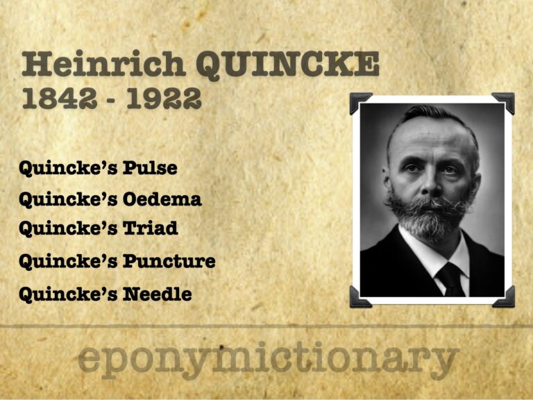
German physician Heinrich Quincke (1842–1922) pioneered lumbar puncture and described Quincke’s pulse, oedema, triad, and more thus shaping modern clinical medicine

Augustus Desiré Waller (1856–1922) was a British physiologist who recorded the first human electrocardiogram (ECG) in 1887. His work laid the foundation for modern electrocardiography and inspired Willem Einthoven’s innovations.
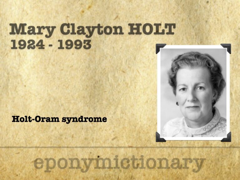
Mary Clayton Holt (1924-1993), English cardiologist. Holt-Oram syndrome (1960); pioneer in cardiac rehab and advocate for women in medicine.
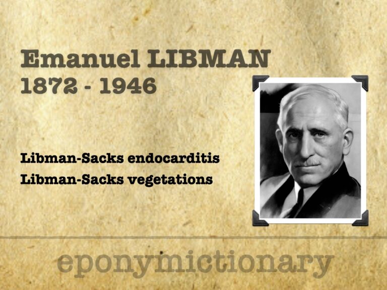
Emanuel Libman (1872–1946), American internist who co-described Libman-Sacks endocarditis and revolutionised diagnostic medicine at Mount Sinai.
François Dessertenne (1917–2001), French cardiologist who coined torsades de pointes in 1966, advanced ECG-based arrhythmia diagnosis with lasting impact.
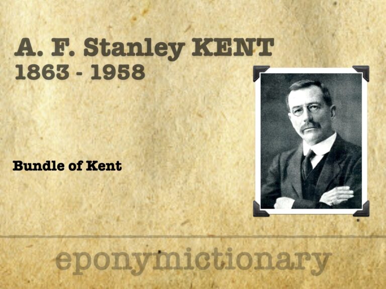
A. F. Stanley Kent (1863–1958), cardiac physiologist; 'bundle of Kent', shaped early electrophysiology; pioneered industrial fatigue science
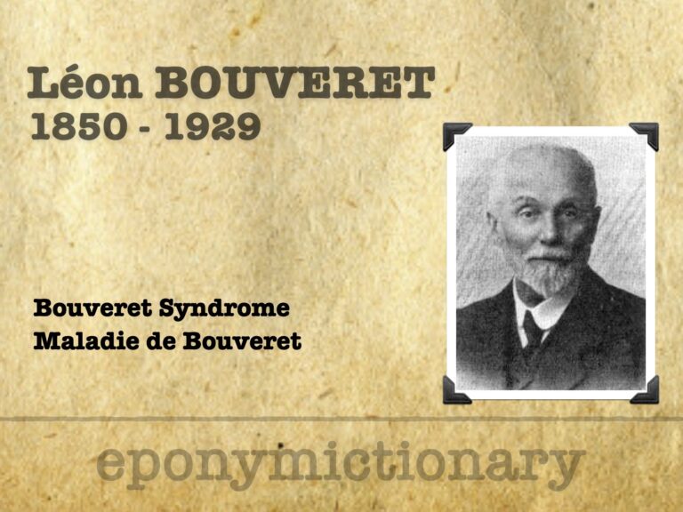
Léon Bouveret (1850-1929) was a French internal medicine physician. Eponymous terms Maladie de Bouveret (1889) and Bouveret Syndrome (1895)
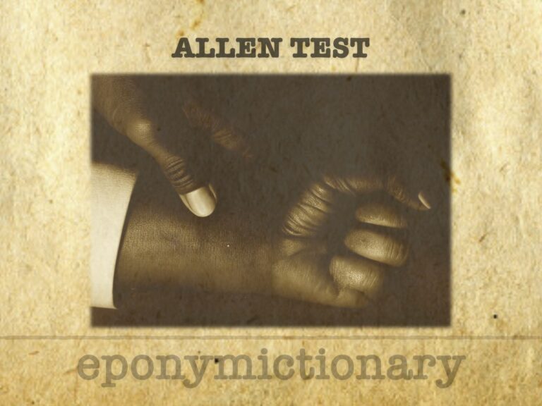
Allen Test: a bedside exam assessing hand arterial flow via radial/ulnar patency; used before ABG, cannulation, or radial artery harvest.
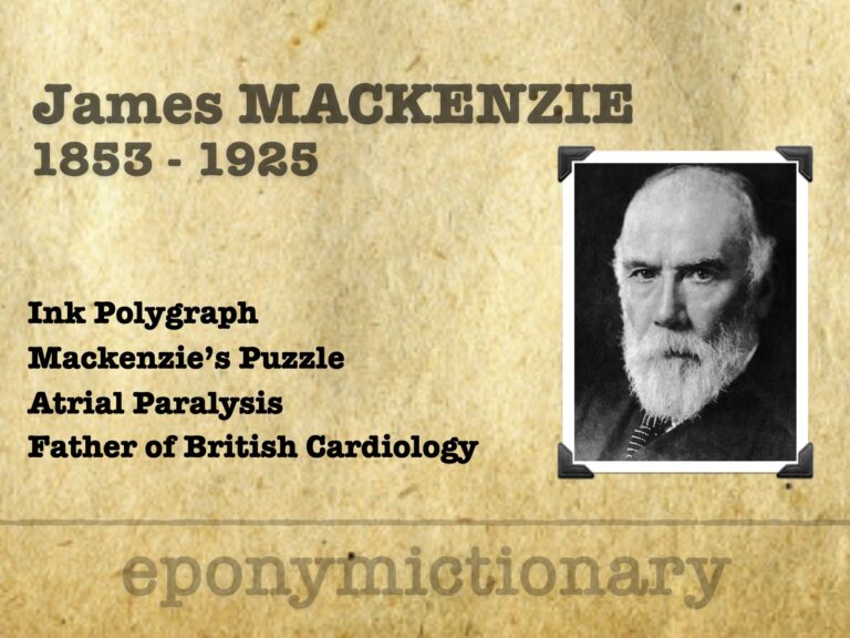
Sir James Mackenzie (1853–1925), Scottish GP and pioneer cardiologist, invented the ink polygraph and defined arrhythmias, angina, and atrial fibrillation
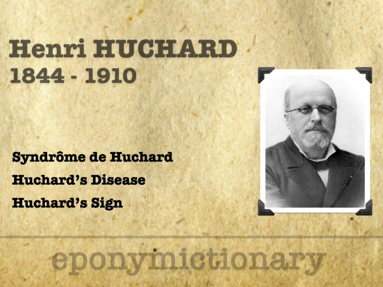
Henri Huchard (1844–1910), French cardiologist at Necker, defined “cardio-arterial” disease, described Huchard’s sign, and helped shape early hypertension care.

Texidor’s Twinge (Precordial Catch Syndrome): benign, sharp chest pain in youth, first described in 1892, clarified by Miller, Texidor, and Asher in the 1950s.