CT Case 004
A 70-year-old female presents with 12 hours duration of headache and progressive drowsiness.
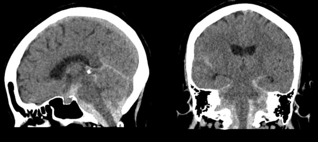
A non-contrast CT scan of the brain is performed.

Describe and interpret the non-contrast images
CT INTERPRETATION (non-contrast)
There is diffuse subarachnoid located within the posterior fossa.
This distribution of blood is unusual for a subarachnoid haemorrhage (SAH).

The diagram below shows the typical location for SAH secondary to berry (saccular) aneurysm rupture.
Normally blood is seen surrounding the Circle of Willis as this is where the majority of berry aneurysms occur

B, CT image representing anatomic structures
Source: Sectional anatomy for radiographers
CLINICAL CORRELATION
This patient has an acute non-traumatic subarachnoid haemorrhage.
A ruptured berry aneurysm is the most common cause of a spontaneous SAH (85% of cases). In this case, the distribution of blood in the posterior fossa suggests an aneurysm or vascular malformation involving the vertebrobasilar system.
Other causes of non-traumatic SAH to consider are perimesenchephalic haemorrhage and amyloid angiopathy.
The CT angiogram for this patient did not show an aneurysm. Instead, it showed an arterio-venous malformation. The appearance is often referred to as a ‘bag of worms’.
Our patient was initially managed with external ventricular drain placement for management of evolving obstructive hydrocephalus. Once stable she underwent successful endovascular repair.
[cite]
TOP 100 CT SERIES
Sydney-based Emergency Physician (MBBS, FACEM) working at Liverpool Hospital. Passionate about education, trainees and travel. Special interests include radiology, orthopaedics and trauma. Creator of the Sydney Emergency XRay interpretation day (SEXI).
Dr Leon Lam FRANZCR MBBS BSci(Med). Clinical Radiologist and Senior Staff Specialist at Liverpool Hospital, Sydney
Emergency Medicine Education Fellow at Liverpool Hospital NSW. MBBS (Hons) Monash University. Interests in indigenous health and medical education. When not in the emergency department, can most likely be found running up some mountain training for the next ultramarathon.


