![]()
CT Case 018
A 55 yo women presents with altered level of consciousness and hypertension.
A CT brain is performed.


Describe and interpret the CT images
CT INTERPRETATION
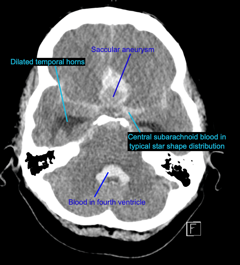
There is a large saccular aneurysm arising at the branching of the anterior communicating artery. It measures 14x20x11mm.
There is surrounding intra-parenchymal haemorrhage in the midline (26x20x26mm) as well as subarachnoid blood in the region of the frontotemporal lobes.
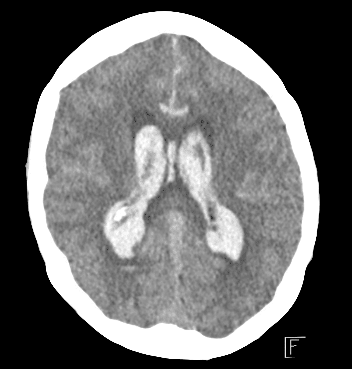
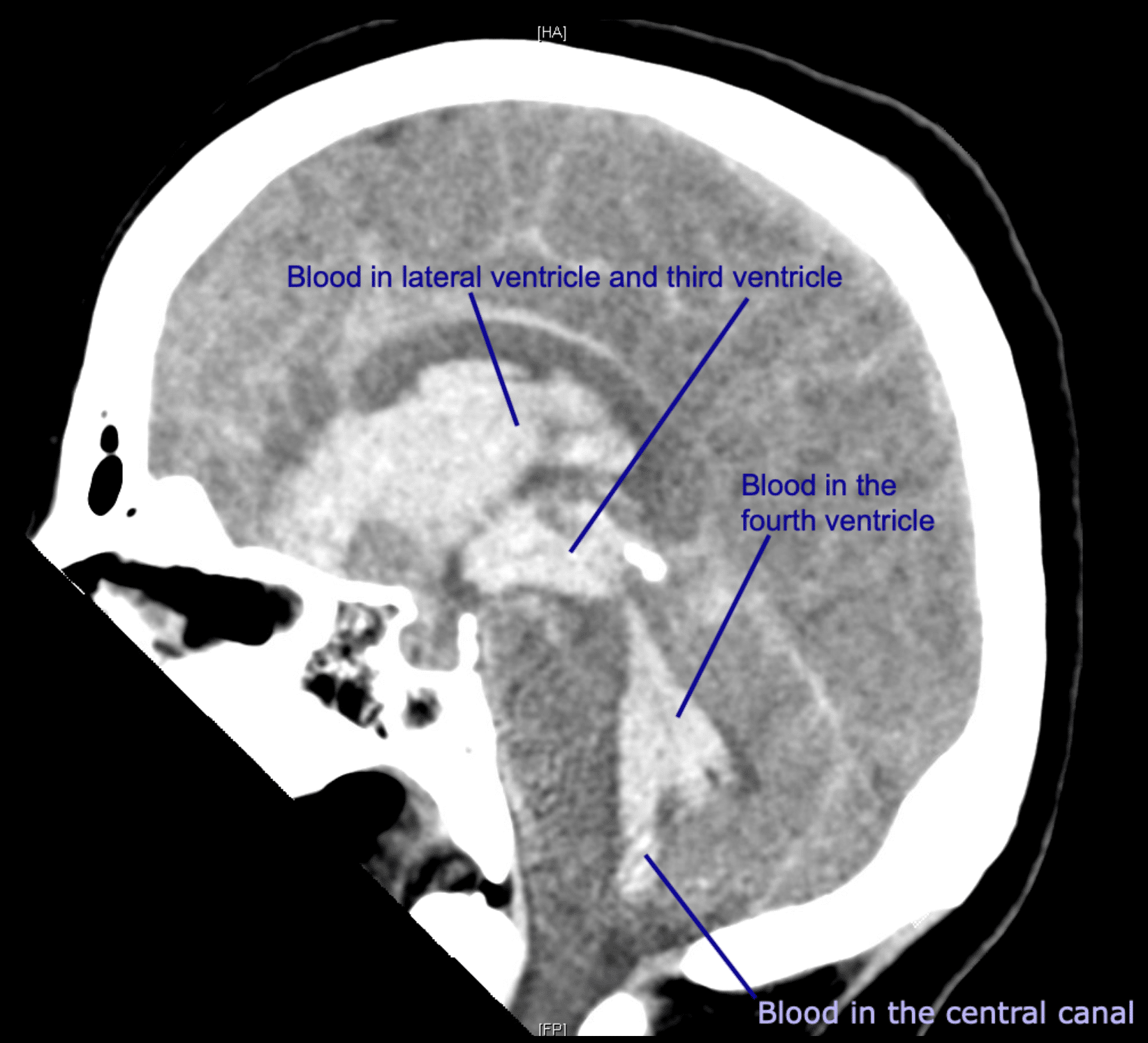
What is even more striking is the extensive haemorrhage in the lateral ventricles, the third and the fourth ventricles and inferiorly in the central canal.

Further imaging
A CT angiogram of the Circle of Willis is then performed
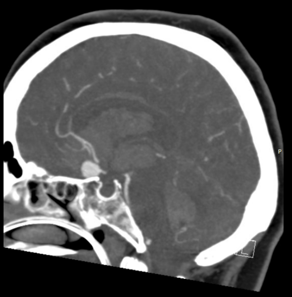

Describe and interpret the CTA images
CT INTERPRETATION


CLINICAL CORRELATION
This is an example of a large anterior communicating artery saccular aneurysm which has ruptured, causing both intra-parenchymal and subarachnoid haemorrhage, and with extension of blood into the ventricles. On CT, when we see dilated temporal horns of the lateral ventricles at this level, as we see in IMAGE 4, it suggests early hydrocephalus.
It is often not possible to see an aneurysm on a non-contrast CT scan. However, the large size of the aneurysm here makes it possible to visualise.
A CT angiogram is still performed for confirmation, to look for other smaller aneurysms and to assist with surgical planning.
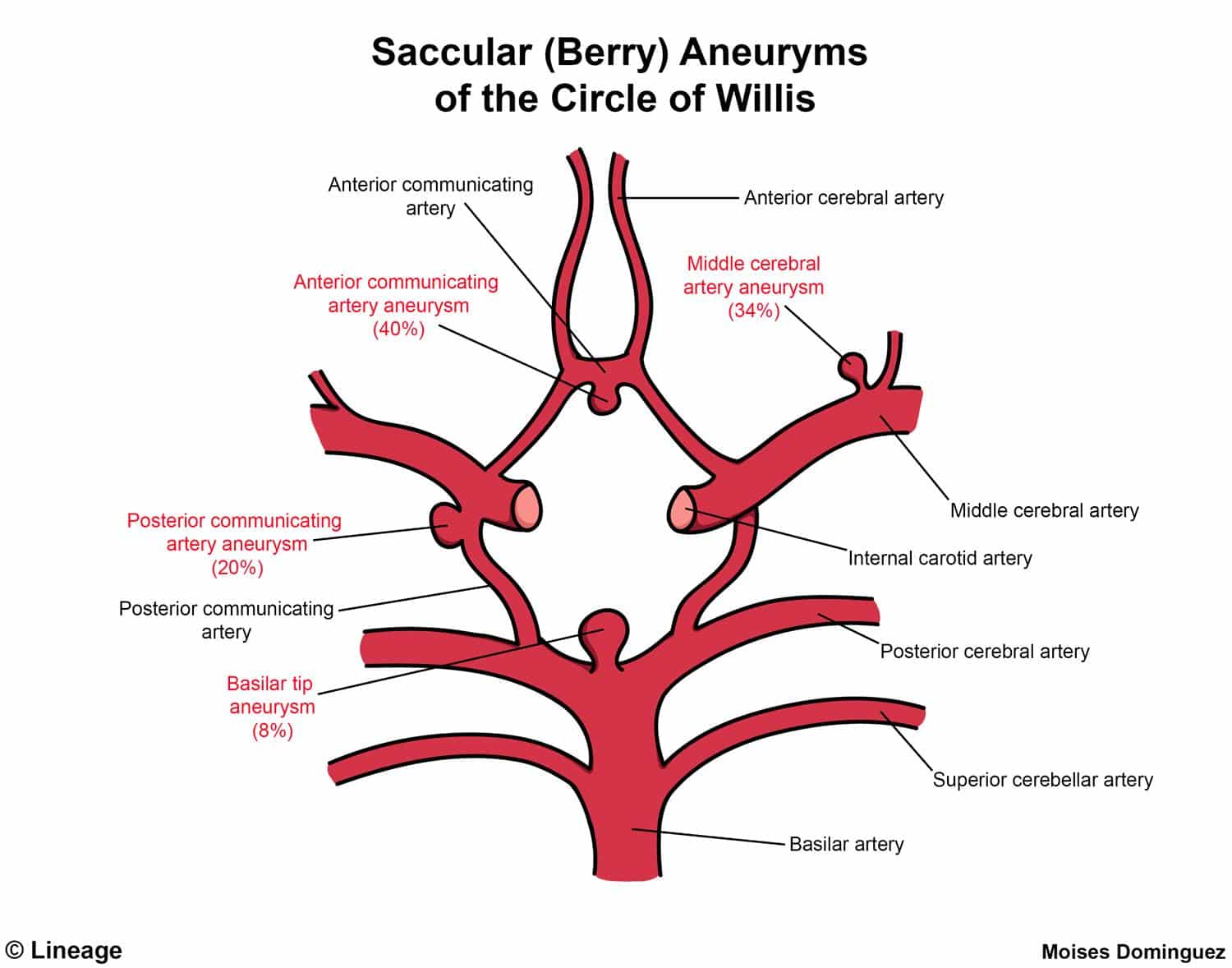
A saccular aneurysm (also known as a berry aneurysm) is the most common cause of non-traumatic SAH. Saccular aneurysms are typical located at the junction between the arteries that make up the Circle of Willis. See diagram below.
While we typically think of ruptured aneurysms causing SAH, they can also cause intra-parenchymal haemorrhage, as occurred in this case.
This is called a ‘jet’ or ‘flame haemorrhage’ given their flame-like appearance. This occurs when an aneurysm abutting the surface of the brain ruptures and the pressure of blood leaving the aneurysm dissects into the brain parenchyma.
This patient was managed with EVD placement followed by surgical repair of the aneurysm.
REFERENCES
- Faluk M, De Jesus O. Saccular Aneurysm. 2022 May 8. In: StatPearls [Internet]
[cite]
TOP 100 CT SERIES

Dr Leon Lam FRANZCR MBBS BSci(Med). Clinical Radiologist and Senior Staff Specialist at Liverpool Hospital, Sydney

Emergency Medicine Education Fellow at Liverpool Hospital NSW. MBBS (Hons) Monash University. Interests in indigenous health and medical education. When not in the emergency department, can most likely be found running up some mountain training for the next ultramarathon.

Sydney-based Emergency Physician (MBBS, FACEM) working at Liverpool Hospital. Passionate about education, trainees and travel. Special interests include radiology, orthopaedics and trauma. Creator of the Sydney Emergency XRay interpretation day (SEXI).