CT Case 024
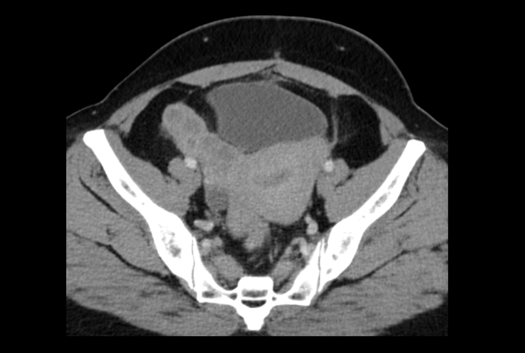
A 48-year-old female presents with 3 days of right lower abdominal pain and fever with a background of previous recurrent pelvic inflammatory disease.


Describe and interpret the CT images
CT INTERPRETATION
The right fallopian tube is filled with fluid, it has a thickened wall and has surrounding fat stranding. In this clinical context this is consistent with pyosalpinx (pus in the fallopian tube).
There is also a right sided complex fluid collection with septations sitting to the right of the uterus, which likely represents an ovarian or paraovarian abscess.
Together these findings are described as a tubo-ovarian abscess, a complication of pelvic inflammatory disease. There is no free fluid in the pelvis to suggest rupture of the abscess.


CLINICAL CORRELATION
Tubo-ovarian abscess is a complication of pelvic inflammatory disease.
The ovaries can often be hard to identify on CT. A trick to identify the ovary is to track back from the gonadal vein towards the ovary. The gonadal vein can be found sitting in front of the iliac veins.
When assessing a case of tubo-ovarian abscess we also need to look for complications.
Complications to look for are abscess rupture (free fluid in the pelvis) and rarely, Fitz-Hugh-Curtis syndrome (peri-hepatitis) which on CT looks like thickening of the liver capsule with perihepatic fluid.
Options for treatment of tubo-ovarian abscess include IV antibiotics, percutaneous drainage, and surgical removal. Several factors (such as presence of sepsis, size of abscess, age of the patient, recurrence of infection) determine the most appropriate treatment. Given the large abscess with loculations surgical management was most appropriate in this case, our patient was managed with right sided oophorectomy and salpingectomy.
References
- Munro K, Gharaibeh A, Nagabushanam S, Martin C. Diagnosis and management of tubo-ovarian abscesses. TOG. 2018; 20 (1): 11-19
[cite]
TOP 100 CT SERIES
Provisional fellow in emergency radiology, Liverpool hospital, Sydney. Other areas of interest include paediatric and cardiac imaging.
Emergency Medicine Education Fellow at Liverpool Hospital NSW. MBBS (Hons) Monash University. Interests in indigenous health and medical education. When not in the emergency department, can most likely be found running up some mountain training for the next ultramarathon.
Dr Leon Lam FRANZCR MBBS BSci(Med). Clinical Radiologist and Senior Staff Specialist at Liverpool Hospital, Sydney
Sydney-based Emergency Physician (MBBS, FACEM) working at Liverpool Hospital. Passionate about education, trainees and travel. Special interests include radiology, orthopaedics and trauma. Creator of the Sydney Emergency XRay interpretation day (SEXI).


