
![]()
CT Case 025
A 50yo normally well man presents with 36 hours of fevers and RLQ pain. He has localised tenderness in McBurney’s point with a pulse rate of 105bpm.
Describe and interpret the CT images
CT INTERPRETATION
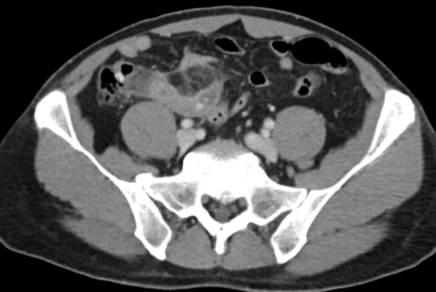
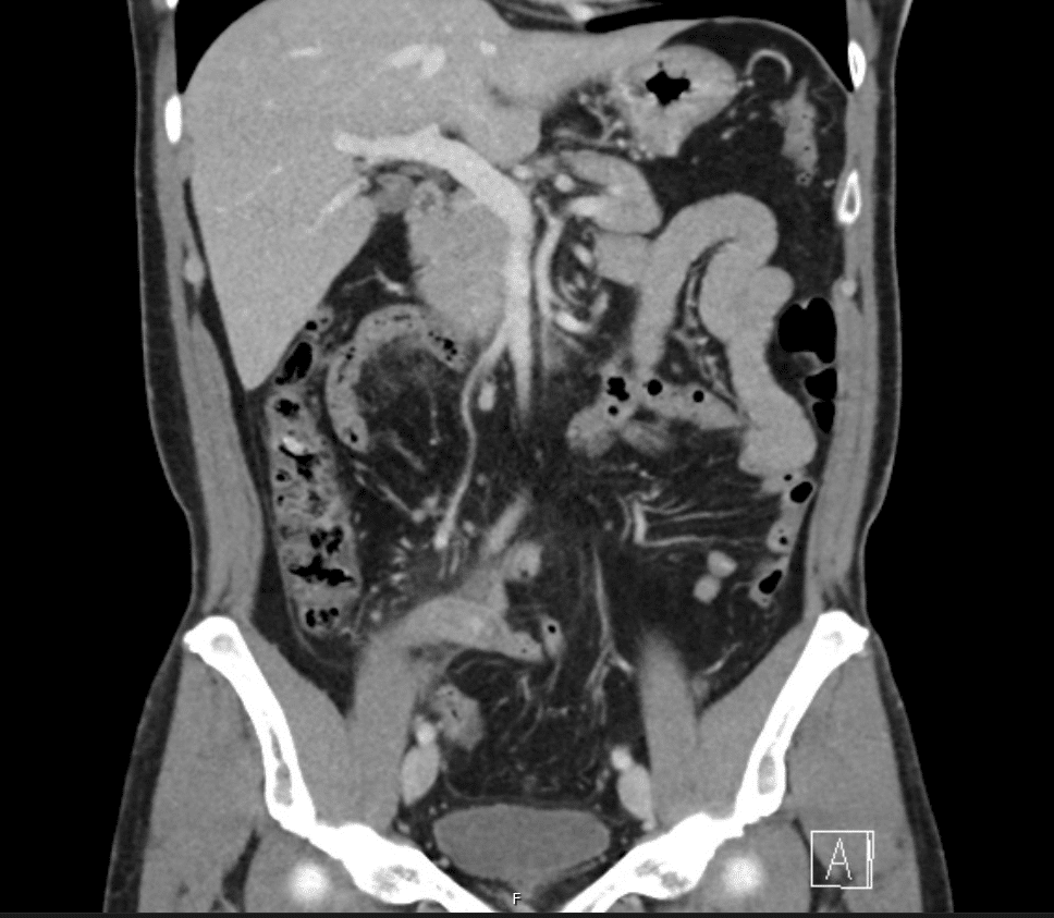
The appendix wall is thickened, the appendix measures up to 11mm in diameter
Appendicoliths are seen within its lumen.
There is marked local fat stranding and trace free fluid.
There is no collection and no pneumoperitoneum.


CLINICAL CORRELATION
This isn’t a trick question!
Here’s an example of plain old appendicitis.
What to look for on CT;
A normal appendix will be surrounded by homogenous, non-inflamed fat and often contains intraluminal gas. Typically, we describe a normal appendix as having a diameter of 6mm or less. However, this is the parameter used for ultrasound scan. In fact, on CT scan, the normal diameter could be up to 8-9mm.
So, in appendicitis we look for;
- A dilated appendix
- Thickening of the wall of the appendix (>3mm) with abnormal increased wall enhancement
- Peri-appendiceal inflammation, suggested by fat stranding and extraluminal fluid
- Lack of gas within the lumen of the appendix
- We might also see local lymph node enlargement and appendicoliths
We also need to look for the complications of appendicitis, namely;
- Abscess
- Perforation (pneumoperitoneum along with free intra-abdominal fluid)
- Gangrene (focal wall non-enhancement)
Note, as we know, the position of the appendix can be very variable. This is what accounts for the variable description of pain that our patients provide. We also need to remember this when trying to find the appendix on CT scan.
Roughly, the position of the appendix is;
- behind the caecum (ascending retrocecal): 65%
- inferior to the cecum (subcaecal): 31%
- behind the cecum (transverse retrocaecal): 2%
- anterior to the ileum (ascending paracaecal preileal): 1%
- posterior to the ileum (ascending paracaecal retroileal): 0.5%

References
- Hartung MP. Abdominal CT: acute appendicitis LITFL
- Connell L, Buttner R. Appendicitis. LITFL
- Cadogan M. Appendicitis – the eponymous examination. LITFL
[cite]
TOP 100 CT SERIES

Emergency Medicine Education Fellow at Liverpool Hospital NSW. MBBS (Hons) Monash University. Interests in indigenous health and medical education. When not in the emergency department, can most likely be found running up some mountain training for the next ultramarathon.

Dr Leon Lam FRANZCR MBBS BSci(Med). Clinical Radiologist and Senior Staff Specialist at Liverpool Hospital, Sydney

Sydney-based Emergency Physician (MBBS, FACEM) working at Liverpool Hospital. Passionate about education, trainees and travel. Special interests include radiology, orthopaedics and trauma. Creator of the Sydney Emergency XRay interpretation day (SEXI).

Provisional fellow in emergency radiology, Liverpool hospital, Sydney. Other areas of interest include paediatric and cardiac imaging.