![]()
CT Case 080
A 70-year-old male presents with 1 week of atraumatic right hip pain.
On examination he has pain on flexion and extension of his right hip and mild tachycardia (HR 105).
He has a past medical history of antiphospholipid syndrome with recurrent pulmonary embolus and on lifelong therapeutic clexane (enoxaparin sodium).


Describe and interpret her CT and MRI scan
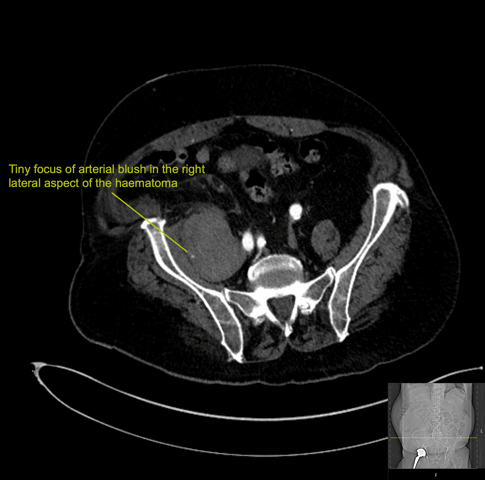
The right psoas muscle is enlarged with hyperdense areas within in keeping with psoas muscle haematoma.
There is a tiny focus of arterial blush in the right lateral aspect of the haematoma.

Clinical Pearls
Psoas muscle haematoma can be either spontaneous or traumatic.
Spontaneous cases may be seen in patients on antiplatelet or anticoagulant therapy, with DIC, or with haemophilia, or may be see in ruptured AAA (case 84).
Traumatic causes may be iatrogenic such as post lumbar surgery or post biopsy.
Patients commonly present with groin or thigh pain. They may also have numbness or paraesthesia and occasionally will have a nerve palsy, with the femoral nerve being most commonly involved.
The incidence of psoas haematomas has slowly increased overtime with the increased use of anticoagulation and antiplatelet therapy. Haemodialysis is also a risk factor.
The incidence of psoas haematoma is 0.6% in elderly patients on anticoagulant therapy.
The mortality rate is surprisingly high (up to 30%).
This patient had a small haemoglobin drop (124 down to 110) and remained haemodynamically stable.
Management involved initial withholding of clexane. Due to ongoing risk of VTE he was then commenced on a heparin infusion as an inpatient.
As there was no further drop in his haemoglobin, clexane was recommenced, and he was discharged home on a reduced dose of clexane.
References
- Seo JG, Yang JC, Kim TW, Park KH. Intramuscular Hematoma on the Psoas Muscle. Korean J Neurotrauma. 2019 Oct 15;15(2):234-238
- Abedini L, Mehrabi S, Hosseinpour R, Jahantab MB, Salehi V, Yavari Barhaghtalab MJ. Non-penetrating traumatic psoas muscle hematoma presenting with gross hematuria: a case report. Int J Emerg Med. 2021 Apr 7;14(1):20.
- Rippey J. Ultrasound Case 074. LITFL
- Hartung MP. Abdominal CT: body wall. LITFL
TOP 100 CT SERIES

Dr Leon Lam FRANZCR MBBS BSci(Med). Clinical Radiologist and Senior Staff Specialist at Liverpool Hospital, Sydney

Sydney-based Emergency Physician (MBBS, FACEM) working at Liverpool Hospital. Passionate about education, trainees and travel. Special interests include radiology, orthopaedics and trauma. Creator of the Sydney Emergency XRay interpretation day (SEXI).

Provisional fellow in emergency radiology, Liverpool hospital, Sydney. Other areas of interest include paediatric and cardiac imaging.

Emergency Medicine Education Fellow at Liverpool Hospital NSW. MBBS (Hons) Monash University. Interests in indigenous health and medical education. When not in the emergency department, can most likely be found running up some mountain training for the next ultramarathon.