![]()
CT Case 094
A 66-year-old lady presents with left sided facial droop and hemiparesis, headache and vomiting.
A CT stroke series is performed
Describe and interpret the CT scans
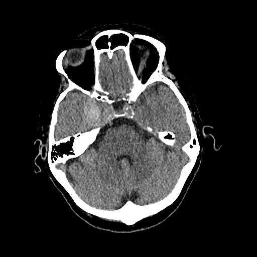
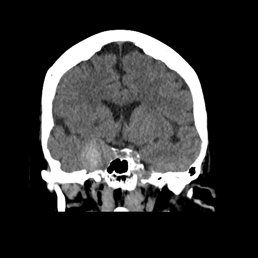
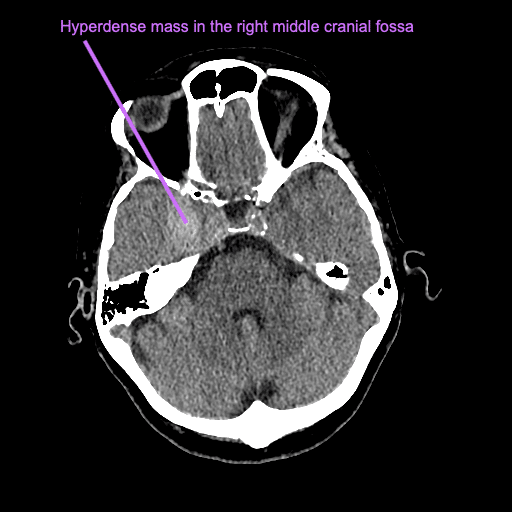
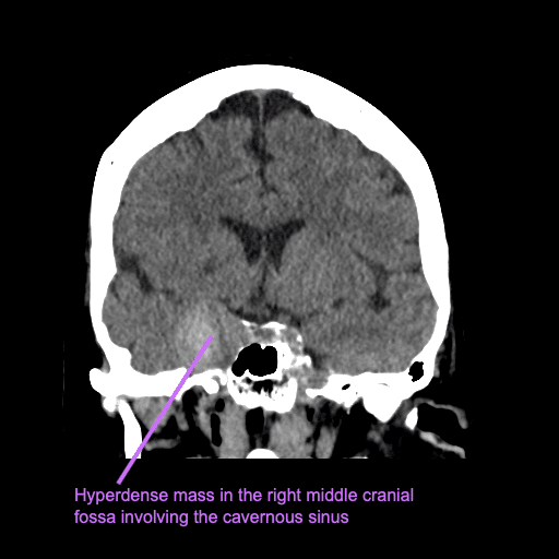
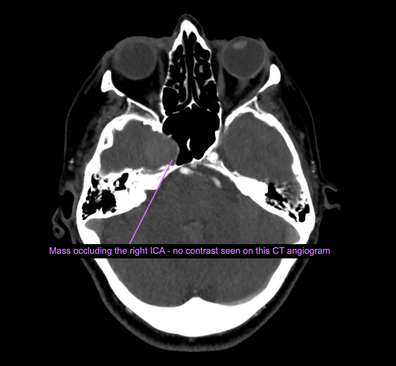
There is a 22mm hyperdense extra-axial mass in the right middle cranial fossa involving the cavernous sinus. It appears that the mass is occluding the right ICA.
There was associated perfusion mismatch in the right ACA/MCA territory.
There is debate about whether the diagnosis is a meningioma or a thrombosed aneurysm. She is commenced on DAPT and planned for further investigation with MRI brain.
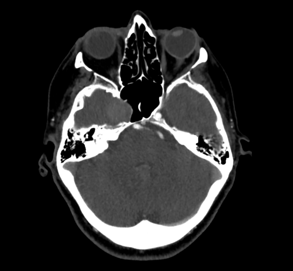
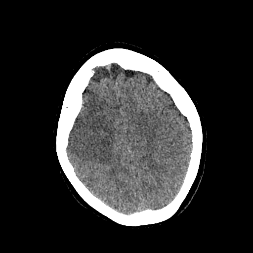
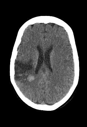
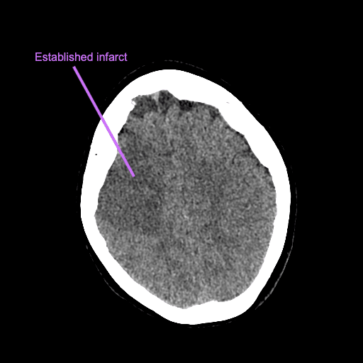
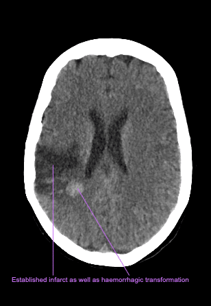
Over the next 48 hours she has progressive symptoms with dysphasia and gaze deviation. She has repeat stroke imaging.
Describe and interpret the CT scans
The subsequent CT showed new perfusion mismatch (not shown here) and a more established infarct, along with haemorrhagic transformation (see CT case 89).
Clinical Pearls
This is a case of a giant intracranial aneurysm with thrombosis.
Giant aneurysms are defined as those greater than 25 mm in diameter.
They make up 5% of all intracranial aneurysms and typically present during the fifth to seventh decades of life.
Unlike in this case, they typically present with mass effect due to their size, they also have a greater risk of rupture (up to 6%), compared to smaller aneurysms.
As in this case, they can also present with ischaemic signs due to thrombus formation with subsequent emboli.
The diagnosis of intracranial aneurysm with thrombus can be difficult to make as the thrombus in the aneurysm can mimic the appearance of a meningioma due to poor intraluminal flow on CT angiogram.
Thrombosed giant aneurysms are also incredibly difficult to manage. The neurointerventional team were consulted in this patient’s care, however there were no neurointerventional options due to the high risk of haemorrhage or further thromboembolic complications.
This patient was continued on DAPT and had ongoing neurorehabilitation.
References
- Borni M, Kolsi F, Cherif I, Boudawara MZ. A giant partial thrombosed aneurysm of the internal cavernous carotid artery mimicking a meningioma of the lesser wing of the sphenoid bone. Radiol Case Rep. 2022 Feb 19;17(4):1325-1329.
- Simon R. Intracranial Mass: Meningioma vs. Thrombosed Aneurysm. Neurology Congress
TOP 100 CT SERIES

Provisional fellow in emergency radiology, Liverpool hospital, Sydney. Other areas of interest include paediatric and cardiac imaging.

Emergency Medicine Education Fellow at Liverpool Hospital NSW. MBBS (Hons) Monash University. Interests in indigenous health and medical education. When not in the emergency department, can most likely be found running up some mountain training for the next ultramarathon.

Dr Leon Lam FRANZCR MBBS BSci(Med). Clinical Radiologist and Senior Staff Specialist at Liverpool Hospital, Sydney

Sydney-based Emergency Physician (MBBS, FACEM) working at Liverpool Hospital. Passionate about education, trainees and travel. Special interests include radiology, orthopaedics and trauma. Creator of the Sydney Emergency XRay interpretation day (SEXI).