![]()
CT Case 098
A 60-year-old female with past history of lung cancer, treated with radiotherapy one year prior, has a routine surveillance CT as an outpatient.
She has a background of CCF with severe LV dysfunction, and right sided dual chamber ICD.
She is recalled to the hospital due to an abnormality on her CT scan. At the time of the phone call, she was doing her Christmas shopping at the local shops. She was asymptomatic and reluctantly returned to the hospital for review.
CT chest
Describe and interpret the CT scan
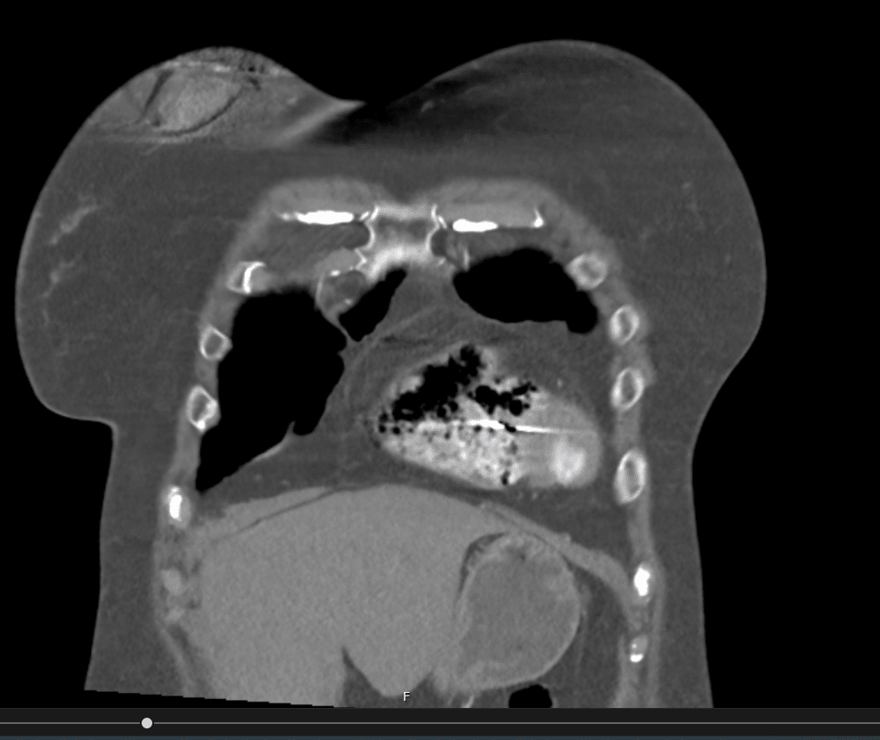
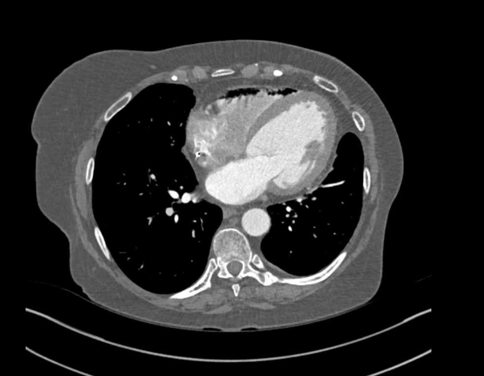
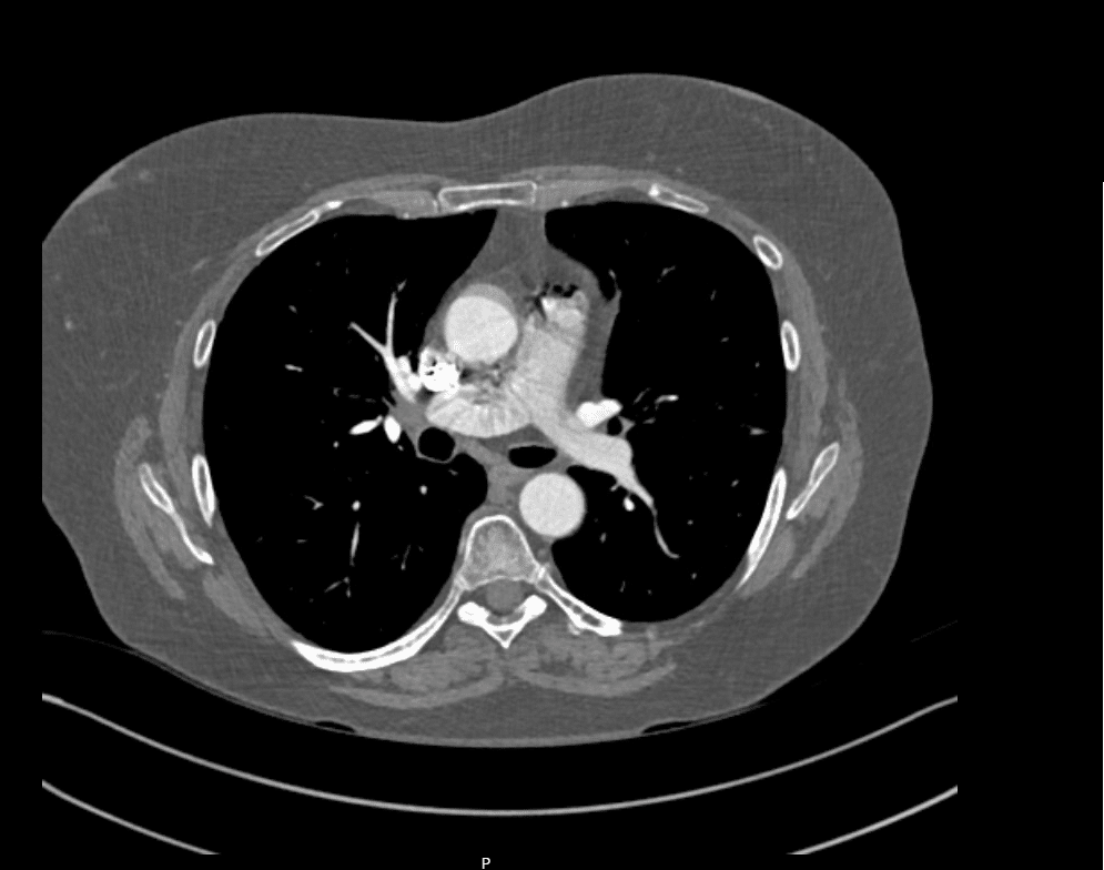
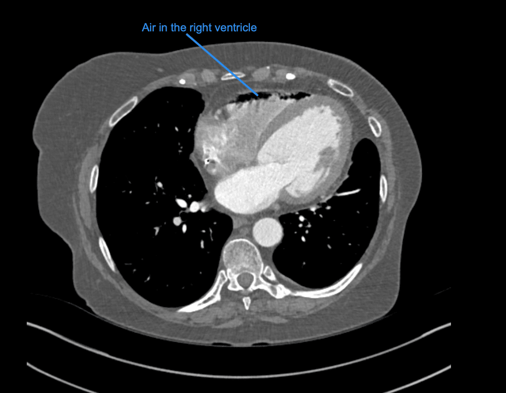
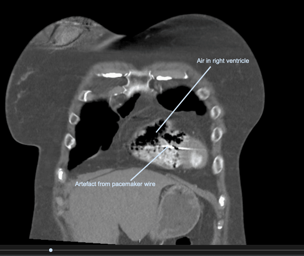
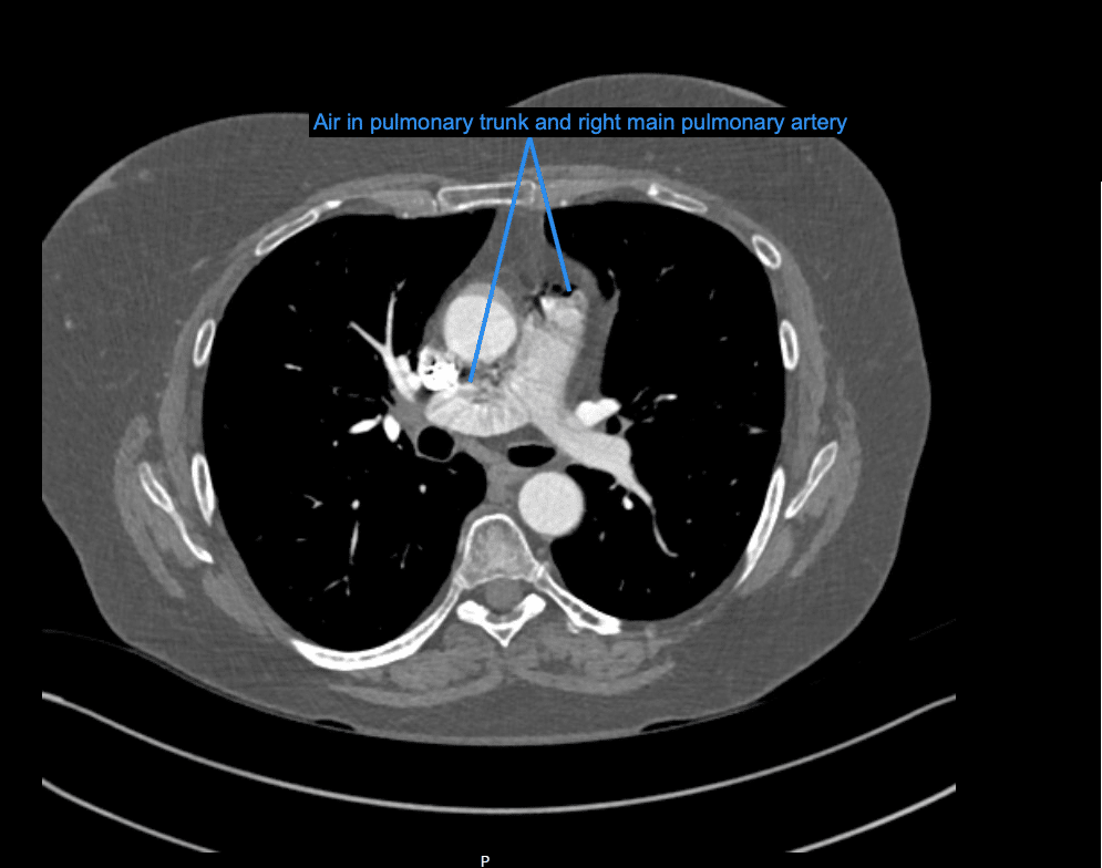
This CT scan demonstrates air embolism.
Within the right ventricle, pulmonary trunk and right pulmonary artery air is seen.
The volume is estimated to be 15-30mL.
Clinical Pearls
Venous gas embolism is defined as an abnormal collection of gas that forms a bubble in the systemic venous system.
It can be related to diving injures, however in most cases it is iatrogenic. In the hospital setting it may be from injection of CT contrast, CVC insertion or removal and haemodialysis. Due to the increase in invasive medical procedures, the incidence is increasing.
Most patients will be asymptomatic. However, when large enough it can affect blood flow and cause haemodynamic instability, dyspnoea or chest pain.
To produce symptoms more than 5ml/kg of gas needs to be introduced into the venous system.
On return to the ED the patient was asymptomatic, with normal sats and no respiratory distress.
Given her unusual presentation, her case was discussed with the local hyperbaric team who recommended 100% oxygen via NRB and repeat CT.
It is believed that the air seen on this patients CT was the result of accidental injection at the time of CT contrast administration.
Treatment of venous gas embolism is largely supportive. 100% oxygen can be used to correct hypoxia as well as to decrease the size of the bubble by a diffusion gradient allowing denitrogenation of the gas embolus. In severe cases hyperbaric oxygen therapy can be considered.
Repeat CT 6 hours later shows complete resolution of gas and she went home to wrap her Christmas presents!
References
- Kerrigan MJ, Cooper JS. Venous Gas Embolism. Stat pearls
TOP 100 CT SERIES

Provisional fellow in emergency radiology, Liverpool hospital, Sydney. Other areas of interest include paediatric and cardiac imaging.

Emergency Medicine Education Fellow at Liverpool Hospital NSW. MBBS (Hons) Monash University. Interests in indigenous health and medical education. When not in the emergency department, can most likely be found running up some mountain training for the next ultramarathon.

Dr Leon Lam FRANZCR MBBS BSci(Med). Clinical Radiologist and Senior Staff Specialist at Liverpool Hospital, Sydney

Sydney-based Emergency Physician (MBBS, FACEM) working at Liverpool Hospital. Passionate about education, trainees and travel. Special interests include radiology, orthopaedics and trauma. Creator of the Sydney Emergency XRay interpretation day (SEXI).