
![]()
CXR Case 006
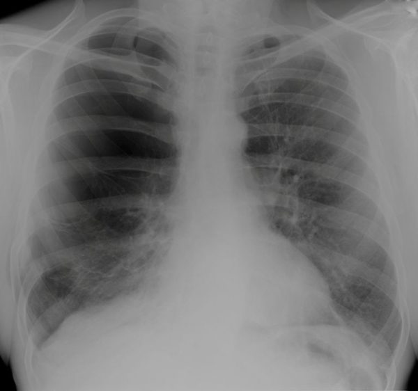
74 yo male ex-smoker presents with worsening breathlessness
Describe and interpret this CXR
CHEST X-RAY INTERPRETATION
There are reduced lung markings throughout the right side with tethers of lung to the chest wall suggesting giant bullae, not pneumothorax
There is a fracture to the right 9th rib, and possibly 8th
CLINICAL CORRELATION
Giant bullae are on the extreme spectrum of emphysema.
They are thought to be more common in marijuana smokers (although are still most commonly seen in emphysema).
The mechanism of dyspnoea in these patients is the same as most COPD – bronchoconstriction (causing wheeze) and gas trapping (leading to loss of functional residual capacity and over-inflation).
CLINICAL PEARLS
A CT chest will be invaluable in delineating between giant bullae and pneumothorax and also guiding chest drain placement if there is a pneumothorax.
Avoid the lung tissue at all costs!
TOP 150 CXR SERIES
![]()
![]()
![]()
Prof Fraser Brims Curtin Medical School, acute and respiratory medicine specialist, immediate care in sport doc, ex-Royal Navy, academic| Top 100 CXR | Google Scholar | ICIS Course ANZ