
![]()
CXR Case 007
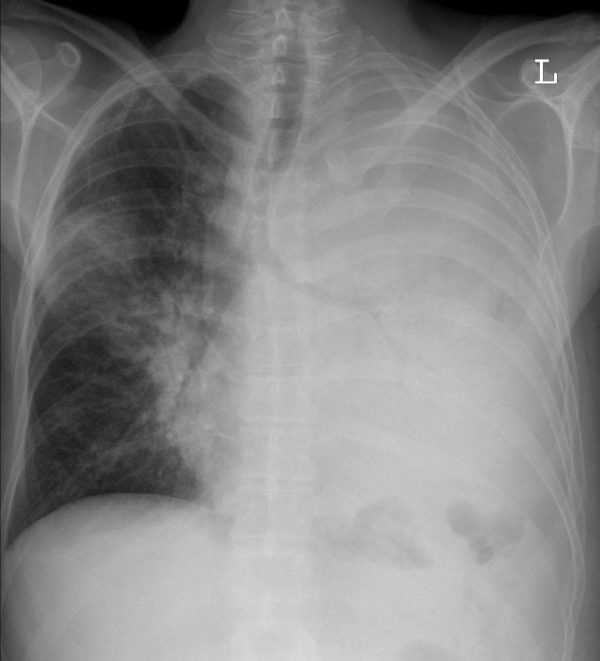
45 year old male presents confused and hypoxaemic
Describe and interpret this CXR
CHEST X-RAY INTERPRETATION
There is complete white out of the left lung with air bronchograms in the mid zone, caused by pneumonia and a smaller patch of consolidation in the right mid zone.
*see CXR case 008 to compare
CLINICAL CORRELATION
There is no collapse in this case.
The trachea and right heart border remain in the normal position.
CLINICAL PEARLS
Severity of pneumonia is mot measured by the degree of consolidation on CXR or CT – its the physiological response to the hypoxaemia and sepsis syndrome that dictate management.
TOP 150 CXR SERIES
![]()
![]()
![]()
Prof Fraser Brims Curtin Medical School, acute and respiratory medicine specialist, immediate care in sport doc, ex-Royal Navy, academic| Top 100 CXR | Google Scholar | ICIS Course ANZ

Isn’t the left side tracheal deviation due to left lung collapse?
The carina and the right heart border show no signs of deviation to the left. I dont think there is volume loss on the left consistent with collapse.