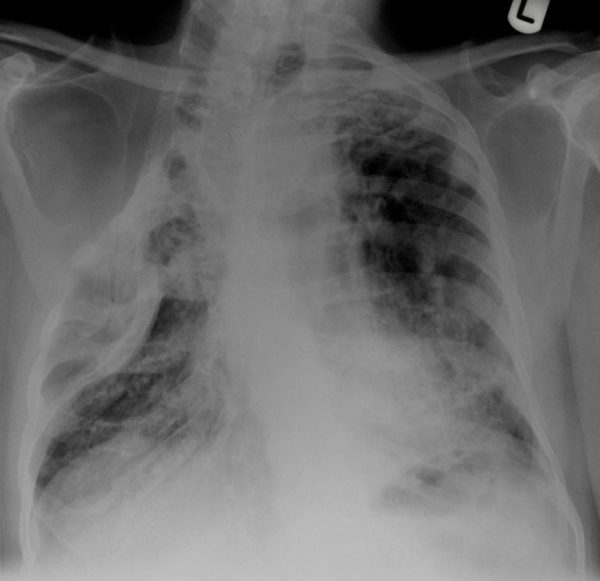
CXR Case 050
An 83 year old man is admitted with acute on chronic type II respiratory failure
Describe and interpret this CXR
CHEST X-RAY INTERPRETATION
The patient has had a thoracoplasty with consequential loss of volume in the right hemithorax.
There is left upper lobe fibrosis.
There is patchy opacification throughout both lung fields likely representing fibrosis however acute infiltrate, particularly in the left lower zone is possible.
*There is marked kyphoscoliosis.
CLINICAL CORRELATION
This man has chronic ventilatory failure secondary to his iatrogenic chest wall deformity
CLINICAL PEARLS
Thoracoplasty – a surgical treatment for the aerobe mycobacterium tuberculosis functionally stops ventilation to the affected upper lobe – and the mycobacteria dies.
Unfortunately significant kyphoscoliosis usually slowly progresses – leading to ventilatory failure.
Prof Fraser Brims Curtin Medical School, acute and respiratory medicine specialist, immediate care in sport doc, ex-Royal Navy, academic| Top 100 CXR | Google Scholar | ICIS Course ANZ

