![]()
CXR Case 071
A 81 year old male presents with progressive cough and occasional wheeze.
click images to enlarge
Describe and interpret this CXR and CT chest
IMAGE INTERPRETATION
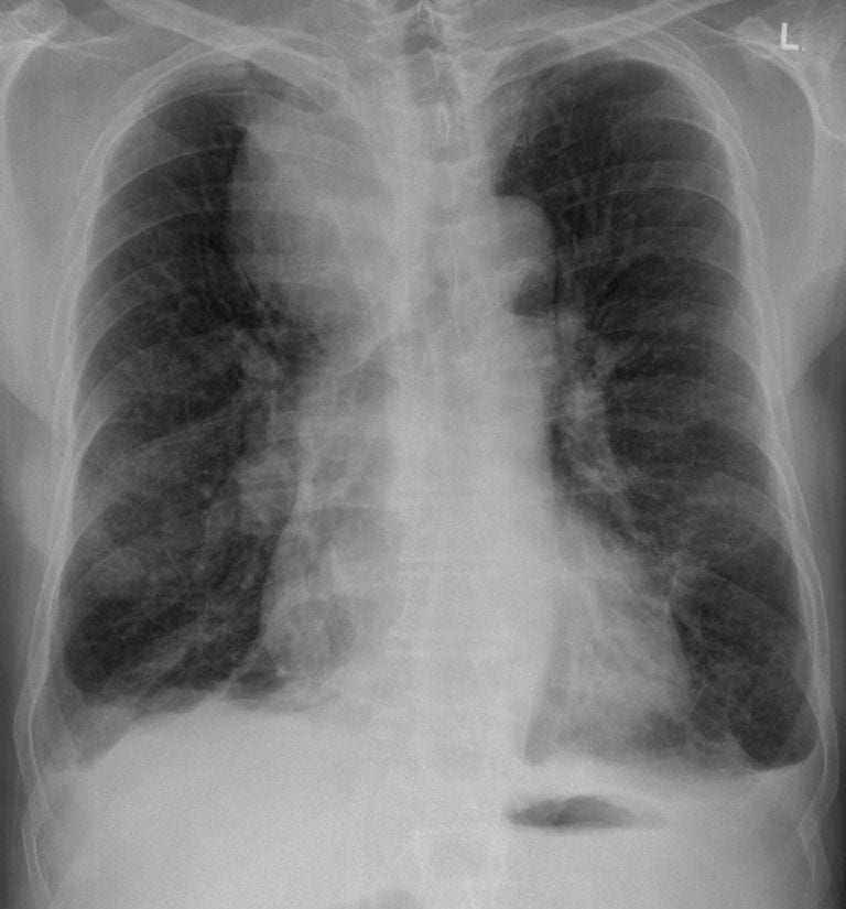
CXR Interpretation:
There is a large soft tissue mass arising from the right superior mediastinum.
The right hilum appears bulky. Both lung fields are over-inflated consistent with COPD.
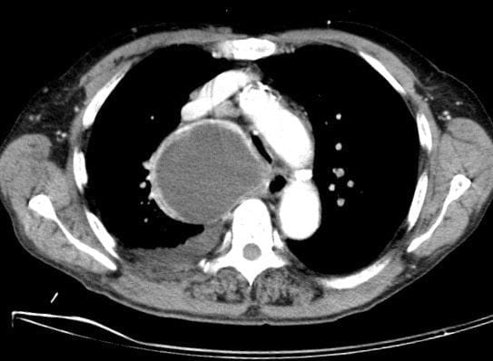
CT Chest Interpretation:
CT demonstrates a large cystic structure arising from the superior mediastinum with thin walls.
It is partially obstructing the airway due to external compression.
CLINICAL CORRELATION
Mediastinal cysts are relatively uncommon.
This will need further work up, however surgery may be indicated.
Causes of mediastinal cysts include bronchogenic, thymic, pericardial, enteric and from teratoma.
CLINICAL PEARLS
While thymic cysts are most common in the anterior mediastinum, others can occur in the middle mediastinum from ectopic remnant thymic tissue
TOP 150 CXR SERIES
![]()
![]()
![]()
Prof Fraser Brims Curtin Medical School, acute and respiratory medicine specialist, immediate care in sport doc, ex-Royal Navy, academic| Top 100 CXR | Google Scholar | ICIS Course ANZ