![]()
CXR Case 079
A 43 yo lady presents with pleuritic chest pain and cough. She is hypotensive, warm and dilated peripherally.
click images to enlarge
Describe and interpret this CXR
CHEST X-RAY INTERPRETATION
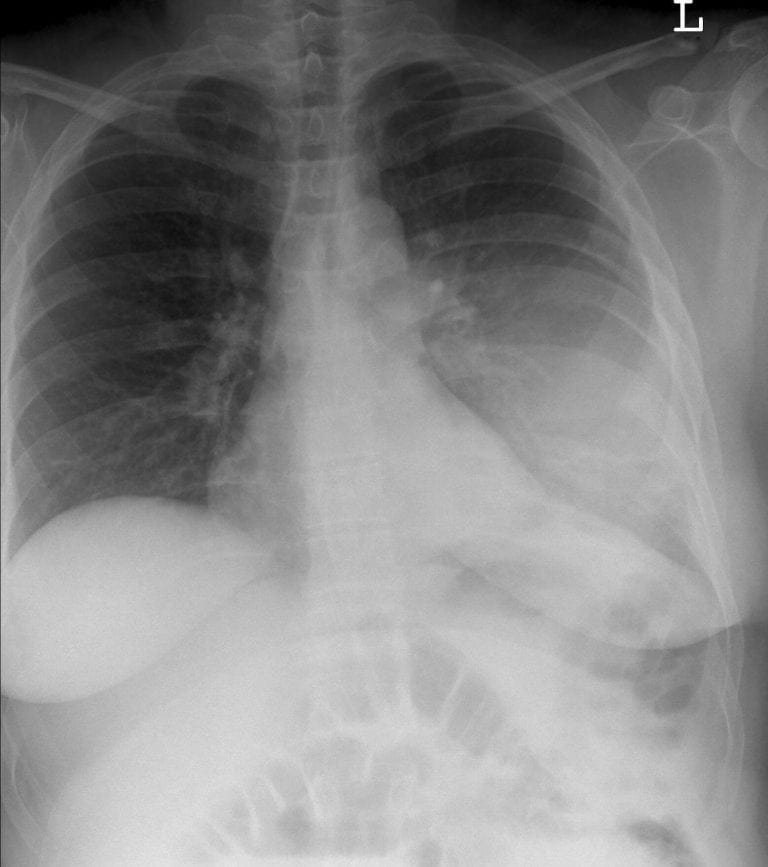
CXR Interpretation:
There is diffuse consolidation in the left lower lung field.
*The left heart border and the diaphragm are maintained (allowing for overlying bowel markings and gas)
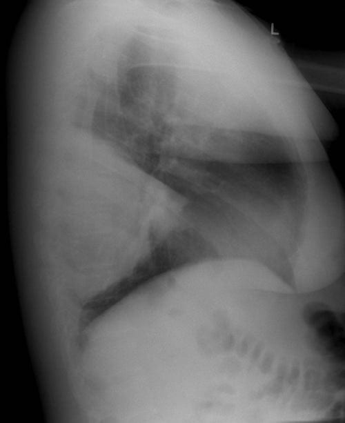
Lateral CXR Interpretation:
This lateral X-ray demonstrated lobar consolidation of the apical segment of the left lower lobe
CLINICAL CORRELATION
This is lobar pneumonia with systemic sepsis.
CLINICAL PEARLS
The radiological appearances of lobar versus more diffuse consolidation do not reliably distinguish between different pathogenic microorganisms.
*While CT chest undeniably offers greater resolution and ability to anatomically define lesions, in most cases it is not routinely indicated over and above the humble CXR.
*Indications for CT in the context of pneumonia would include concern with possible complications, such as development of parapneumonic effusion, haemoptysis or cavitation.
TOP 150 CXR SERIES
![]()
![]()
![]()
Prof Fraser Brims Curtin Medical School, acute and respiratory medicine specialist, immediate care in sport doc, ex-Royal Navy, academic| Top 100 CXR | Google Scholar | ICIS Course ANZ