![]()
CXR Case 086
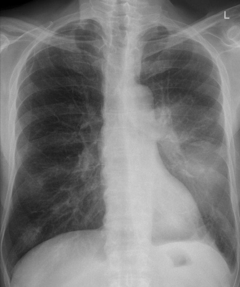
A 34 year old man is admitted with 10 days of worsening fever, malaise and chest pain.
click images to enlarge
Describe and interpret this CXR
CHEST X-RAY INTERPRETATION
There is a diffuse hazy shadowing over the lower left lung field.
There is loss of volume in the left lower hemithorax (right heart border shifted to the left)
Appearances suggest a loculated posterior-basal effusion.
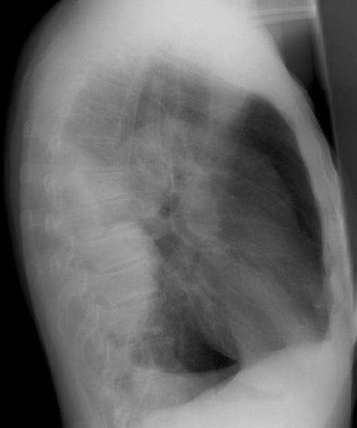
Lateral X-ray demonstrates a large multi-loculated posterior and basal pleural effusion.
CLINICAL CORRELATION
This man has a pleural empyema.
Broad spectrum intravenous antibiotics to cover community acquired pneumonia are required, unless there is recent hospitalization which could change the possible pathogen profile.
CLINICAL PEARLS
The role of intrapleural fibrinolytics is still not certain, although intrapleural tPa with DNAse may have good outcomes in some.
(Early) Video Assisted Thorascopic Surgery (VATS) to clear adhesions and wash the pleural space has a clear role in those who are fit enough.
TOP 150 CXR SERIES
![]()
![]()
![]()
Prof Fraser Brims Curtin Medical School, acute and respiratory medicine specialist, immediate care in sport doc, ex-Royal Navy, academic| Top 100 CXR | Google Scholar | ICIS Course ANZ