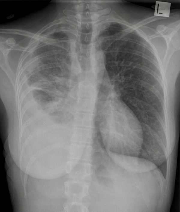
CXR Case 094
44 year old lady admitted with worsening dyspnoea, right sided chest pain, haemoptysis and weight loss of 10kgs in 8 weeks.
Describe and interpret this CXR
CHEST X-RAY INTERPRETATION
There is a partially loculated right pleural effusion. There are increased reticulonodular markings throughout the left lung field.
Left pleural space clear, bones appear normal.
CLINICAL CORRELATION
This is a primary lung cancer with malignant pleural effusion on the right and lymphangitis carcinomatosa on the left side
CLINICAL PEARLS
Lymphangitis carcinomatosa / carcinomatosis is a malignant infiltration of the pulmonary lymphatics.
Frequently associated with lung cancer, but also breast, stomach, pancreatic and prostate.
Oral corticosteroids and diuretics can have transient benefit, but usually this is part of a rapid decline.
Prof Fraser Brims Curtin Medical School, acute and respiratory medicine specialist, immediate care in sport doc, ex-Royal Navy, academic| Top 100 CXR | Google Scholar | ICIS Course ANZ

