
![]()
CXR Case 100
A 39 year old man presents with sudden onset right sided pleuritic pain. He smokes marijuana. SpO2 on air 96%
Describe and interpret this CXR
CHEST X-RAY INTERPRETATION
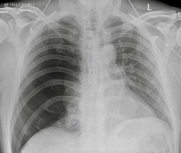
There is a large right sided pneumothorax.
No evidence of trauma.
The left lung looks congested but no obvious parenchymal disease.
CLINICAL CORRELATION
With no known underlying lung disease this is a primary spontaneous pneumothorax (PSP; although the fact that a pneumothorax has occurred suggests underlying lung disease…).
Marijuana smoking is associated with the development of early bullous emphysema, although this is not apparent on the left side.
CLINICAL PEARLS
The size measurement of PSP is arbitrary
- the British guidelines suggest >2cm from chest wall to pleural edge at the level of the hilum
- the Americans suggest >3cm from apex to cupola.
Neither matter very much – the increasing evidence is that PSP can either be left completely alone and will resolve slowly, or simple aspiration using a 14G or 16G cannula is all that’s needed.
The absolute ‘size’ of the pneumothorax doesn’t really matter.. although if you feel the need to determine the exact volume, the most accurate means of assessment is with a CT scan.
TOP 150 CXR SERIES
Prof Fraser Brims Curtin Medical School, acute and respiratory medicine specialist, immediate care in sport doc, ex-Royal Navy, academic| Top 100 CXR | Google Scholar | ICIS Course ANZ