
![]()
CXR Case 106
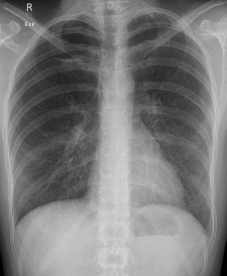
54 year old man presents with worsening breathlessness over 2 days. He is a current smoker and has had periodic breathlessness and wheeze for 4 or 5 years.
Describe and interpret this CXR
CHEST X-RAY INTERPRETATION
There is a small right pneumothorax in the right upper zone.
The lung parenchyma looks normal, pleural spaces are clear.
CLINICAL CORRELATION
This is a spontaneous secondary pneumothorax (SSP) from COPD.
There is no clear evidence base to inform clinical decision making in this situation.
- No intervention (allow time to resolve once visceral pleural defect healed) is possible but a bit controversial in SSP.
- Simple needle aspiration is relatively safe and may be effective.
- Placement of a drain may be dangerous as the pneumothorax is relatively small.
CLINICAL PEARLS
A CT chest is the best way of imaging a pneumothorax – and can guide the safest site for intervention, should this be necessary.
TOP 150 CXR SERIES
Prof Fraser Brims Curtin Medical School, acute and respiratory medicine specialist, immediate care in sport doc, ex-Royal Navy, academic| Top 100 CXR | Google Scholar | ICIS Course ANZ