![]()
CXR Case 111
A 55 year old man is transferred to emergency following a collapse at a sporting event.
Paramedics were unable to record oxygen saturation on scene, a bystander thinks there was a pulse throughout.
Describe and interpret this CXR
CHEST X-RAY INTERPRETATION
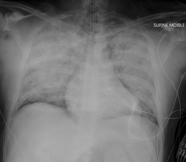
There is widespread, bilateral upper lobe predominant airspace opacification.
Heart size appears normal.
An ETT is present and in a satisfactory position.
CLINICAL CORRELATION
This man has acute pulmonary oedema.
ECG demonstrated (presumed new) LBBB.
Bedside echo confirmed a hypokinetic and very poor LV function in the LAD territory and he was taken to the cardiac catheterization lab.
CLINICAL PEARLS
The air bronchograms visible in the medial area of the upper lobes represent airways that are not full of fluid, surrounded by alveoli and bronchioles that are full of fluid.
Most commonly air bronchograms are associated with pneumonia.
TOP 150 CXR SERIES
Prof Fraser Brims Curtin Medical School, acute and respiratory medicine specialist, immediate care in sport doc, ex-Royal Navy, academic| Top 100 CXR | Google Scholar | ICIS Course ANZ