![]()
CXR Case 112
A 77 yo old lady with a history of ischaemic heart disease is admitted to the ER with symptoms of increasing breathlessness and cough.
Describe and interpret this CXR
CHEST X-RAY INTERPRETATION
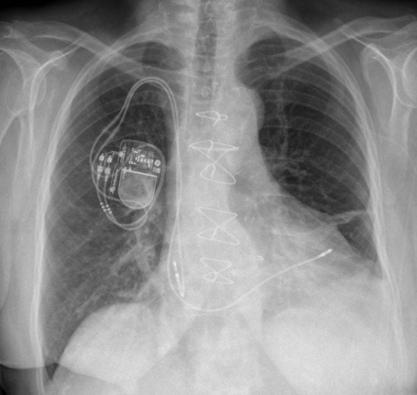
There is a possible left pleural effusion or pleural thickening with a band of atelectasis in the left base.
There may be bronchial wall thickening in the left lower lobe behind the heart.
The rest of the lung parenchyma is clear.
There is a dual chamber PPM and wires from a sternotomy.
There is also a metallic AVR.
CLINICAL CORRELATION
This lady had bronchitis relating to some mild bronchiectasis, and chronic pleural thickening, presumably from old infection.
CLINICAL PEARLS
Note the subtle appearance of the AVR melding with the more prominent sternotomy wires. An important reason to have a clear sequence when reading a chest film…and also not to be distracted by the big obvious stuff.
TOP 150 CXR SERIES
Prof Fraser Brims Curtin Medical School, acute and respiratory medicine specialist, immediate care in sport doc, ex-Royal Navy, academic| Top 100 CXR | Google Scholar | ICIS Course ANZ