
![]()
CXR Case 118
An 81 year old man is found hypoxaemic and confused by paramedics.
click images to enlarge
Describe and interpret this CXR
CHEST X-RAY INTERPRETATION
CXR Interpretation:
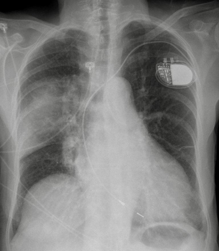
There is airspace shadowing in the right upper lobe with some volume loss.
The right hemidiaphragm is raised anteriorly.
There is a single chamber pace maker in situ.
Left lung field appears hyper-inflated consistent with air trapping from COPD.
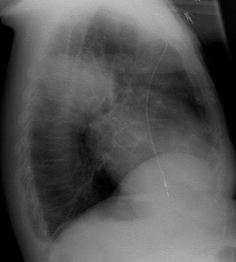
Lateral CXR Interpretation:
The lateral CXR confirms consolidation within the posterior segment right upper lobe.
CLINICAL CORRELATION
Common things are common, so this is likely to be pneumonia complicating COPD.
Given the possible raised diaphragm and proximity of the air space shadowing to the hilum / mediastinum a more proximal lung cancer picking off the right phrenic nerve and causing obstructive pneumonia is possible.
If you want to get very carried away, then the confusion could be from brain metastases … but confusion from hypoxaemia and sepsis is most likely.
CLINICAL PEARLS
CT would be indicated to answer the ?bronchogenic cancer question.
The timing should depend on how worried clinically you are – if it all fits with infection then repeat imaging in ~8 weeks is OK, if increased suspicion (eg additional weight loss, haemoptysis, clubbing) then CT sooner is indicated.
But remember that in the presence of consolidation from infection it might be hard to categorically say there is not something there…
TOP 150 CXR SERIES
Prof Fraser Brims Curtin Medical School, acute and respiratory medicine specialist, immediate care in sport doc, ex-Royal Navy, academic| Top 100 CXR | Google Scholar | ICIS Course ANZ