![]()
CXR Case 119
A 67 year old lady is admitted from respiratory clinic with cough and haemoptysis. She is a never-smoker.
click images to enlarge
Describe and interpret this CXR
CHEST X-RAY INTERPRETATION
CXR Interpretation:
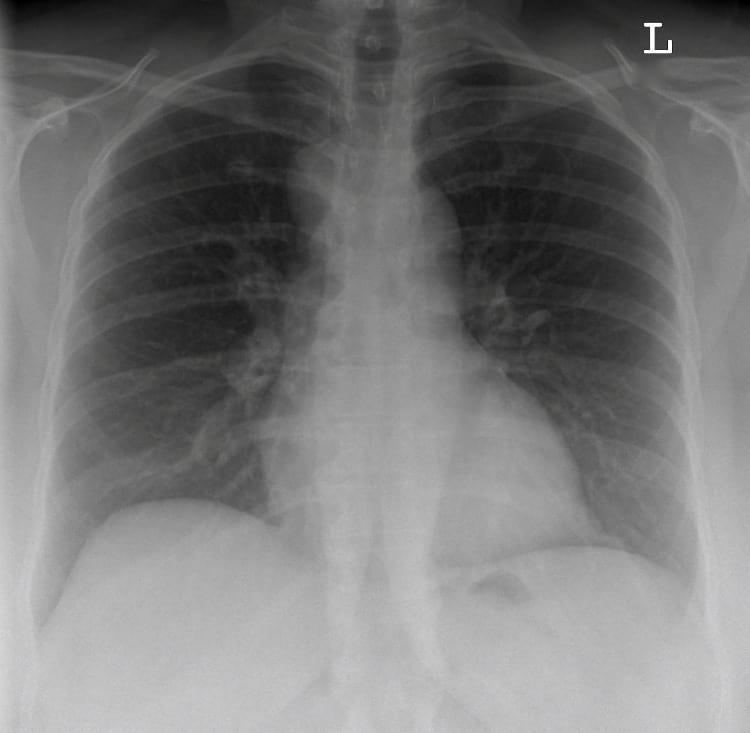
There is a right sided mass in the superior mediastinum with loss of clarity of the tracheal border.
Right hilum is possibly bulky.
Parenchyma and pleura are clear. Bones normal.
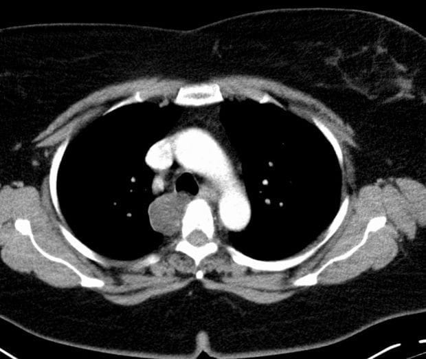
CT Chest Interpretation:
CT chest demonstrates a posterior-superior mediastinal mass, infiltrating the membranous tracheal wall.
CLINICAL CORRELATION
This is a non-small cell lung cancer (NSCLC).
After diagnostic bronchoscopy this lady needs (semi) urgent radiotherapy for the haemoptysis.
CLINICAL PEARLS
Lung cancer in never-smokers is slowly becoming more common, particularly in women.
In some areas within Asia up to 30% of NSCLC in women is in never-smokers.
TOP 150 CXR SERIES
Prof Fraser Brims Curtin Medical School, acute and respiratory medicine specialist, immediate care in sport doc, ex-Royal Navy, academic| Top 100 CXR | Google Scholar | ICIS Course ANZ