
![]()
CXR Case 129
A 29 year old man with a history of deafness presents with a self terminating seizure.

click images to enlarge
Describe and interpret this CXR and CT chest
IMAGE INTERPRETATION
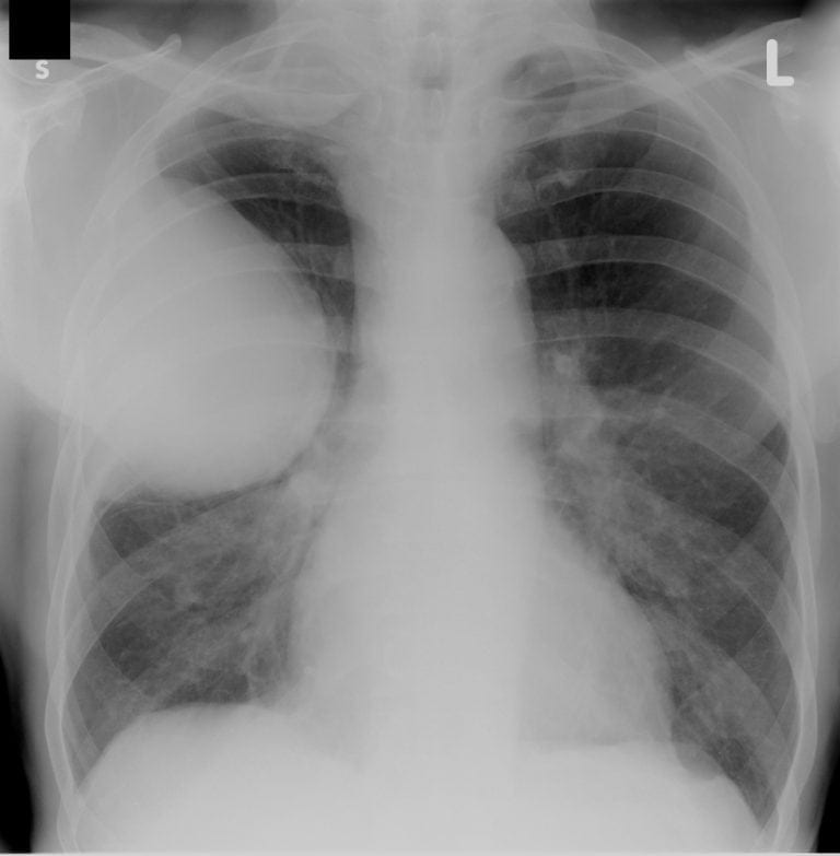
CXR Interpretation:
There is a large rounded pleural based opacity in the right upper zone of the chest.
*There is also increased opacity in the right lung apex, behind the clavicle
CT Chest Interpretation:
CT chest demonstrates a large heterogeneous pleural based mass in the right hemithorax.
There is no mediastinal adenopathy.
CLINICAL CORRELATION
MRI brain demonstrated multiple meningiomas and bilateral acoustic neuromas.
*This is neurofibromatosis type II
*The lesions on the CXR are large schwannomas
CLINICAL PEARLS
Large peripheral schwannomas are unusual features of NF type II
TOP 150 CXR SERIES
Prof Fraser Brims Curtin Medical School, acute and respiratory medicine specialist, immediate care in sport doc, ex-Royal Navy, academic| Top 100 CXR | Google Scholar | ICIS Course ANZ