
![]()
ECG Case 015
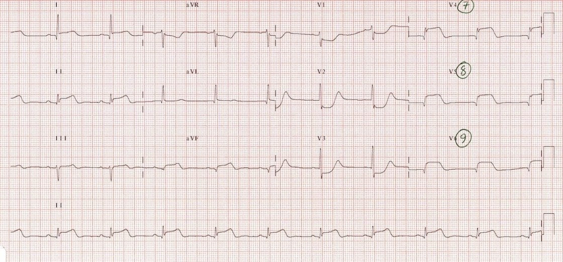
Middle aged patient presenting with central chest pain. Posterior leads V7-9. What does the ECG show?
Describe and interpret this ECG
ECG ANSWER and INTERPRETATION
This is the same patient from ECG 014
Posterior leads confirm the presence of posterior wall infarction by demonstrating typical STEMI morphology:
- ST elevation in V7-9
- Q waves in V7-9
- Inversion of the terminal portion of the T wave (“U wave inversion“) in V7-9
CLINICAL PEARLS
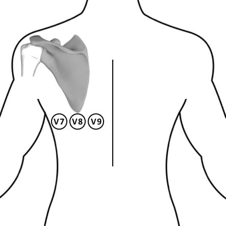
How To Record Posterior Leads
Simply move the V4-6 electrodes around to the back in the same horizontal plane as V6. Annotate the ECG accordingly.
Approximate positions for V7-9 are:
- V7 – left posterior axillary line
- V8 – tip of left scapula
- V9 – left paraspinal region
References
Further Reading
- Wiesbauer F, Kühn P. ECG Mastery: Yellow Belt online course. Understand ECG basics. Medmastery
- Wiesbauer F, Kühn P. ECG Mastery: Blue Belt online course: Become an ECG expert. Medmastery
- Kühn P, Houghton A. ECG Mastery: Black Belt Workshop. Advanced ECG interpretation. Medmastery
- Rawshani A. Clinical ECG Interpretation ECG Waves
- Smith SW. Dr Smith’s ECG blog.
- Wiesbauer F. Little Black Book of ECG Secrets. Medmastery PDF
TOP 100 ECG Series
Emergency Physician in Prehospital and Retrieval Medicine in Sydney, Australia. He has a passion for ECG interpretation and medical education | ECG Library |
This was really cool to see the continuation of the previous case