ECG Case 087
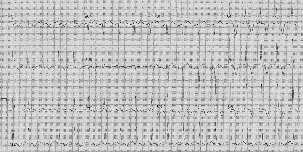
36yr old male presented to the Emergency Department with a non-cardiac issue but an ECG was performed due to a pre-existing cardiac history.
Describe and interpret this ECG
ECG ANSWER and INTERPRETATION
Rate:
- ~115 bpm
Rhythm:
- Regular
- Sinus Rhythm
Axis:
- Normal
Intervals:
- PR – Normal (~160ms)
- QRS – Normal (100ms)
- QT – 320ms
Segments:
- ST elevation leads aVR (<1mm), V1 (1mm), V2 (2mm)
- ST depression lead I
Additional:
- Biphasic T wave leads aVF & V3
- T wave inversion leads I, II, aVL and deep inversion leads V4-6
- Voltage criteria for LVH
- S V2 (25mm) + R V6 (20mm)
- R wave peak time in leads V5 & V6 ~60ms
- P waves wide with deep terminal deflection in lead V1 with notching in lead II
Interpretation:
- Sinus tachycardia
- ECG features for:
- LVH
- LAE
- Deep anterolateral T wave inversion
Broad differentials
These ECG features could be seen in a wide range of conditions and include:
- Ischaemia
- Myocarditis
- Acid-base / electrolyte disturbance
- Raised ICP
OUTCOME
The key to interpretation of any ECG, or any test for that matter, is looking at the test and then taking it back to the patient in question.
In this case we have a young male with no acute cardiac complaint, i.e. nil chest pain, dyspnea, and a known cardiac condition. Given these factors the most likely cause is apical hypertrophic cardiomyopathy (AHCM) given the absence of Q waves and deep T wave inversion in the precordial leads, this patient did in fact have known apical hypertrophic cardiomyopathy.
You can read more about apical hypertrophic cardiomyopathy in the links below:
- ECG Library – Hypertrophic Cardiomyopathy (HCM)
- Madias JE. Electrocardiogram in apical hypertrophic cardiomyopathy with a speculation as to the mechanism of its features. Neth Heart J. 2013 Jun;21(6):268-71. [PMC3661871]
- Yıldırım MN, Selçoki Y, Eryonucu B. Apical Hypertrophic Cardiomyopathy. Eur J Gen Med 2010;7(2):206-209
TOP 100 ECG Series
Emergency Medicine Specialist MBChB FRCEM FACEM. Medical Education, Cardiology and Web Based Resources | @jjlarkin78 | LinkedIn |

