


Gunnar Wiberg
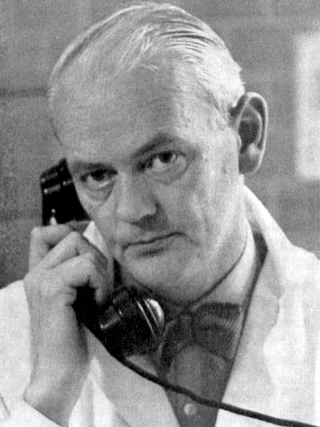
Georg Gunnar Fredrik Wiberg (1902–1988) was a Swedish orthopaedic surgeon, academic and clinical researcher best known for introducing the centre-edge (CE) angle.
Trained at the Karolinska Institute and later professor of orthopaedics at Lund University, Wiberg made important contributions to the study of hip dysplasia, slipped capital femoral epiphysis, patellofemoral anatomy and chondromalacia, as well as the mechanisms of pain in intervertebral disc disease. His 1939 thesis linked acetabular dysplasia and congenital subluxation with later osteoarthritis and introduced the centre-edge angle of Wiberg, which remains widely used in the assessment of lateral acetabular coverage.
Wiberg also helped shape modern Swedish orthopaedics, particularly the transfer of acute fracture care from general surgery to orthopaedic services. Internationally respected, he was fluent in English, German and French and became an honorary member of several foreign orthopaedic societies.
Outside medicine he was an accomplished tennis player and skilled woodworker. He designed wooden operating-theatre clogs and the logotype of the Scandinavian Orthopaedic Association. His successor Göran Bauer remembered him as “a pioneer of orthopedics and as a gentleman.”
Biographical Timeline
- Born November 24, 1902 in Göteborg (Gothenburg), Sweden
- 1921 – Completed secondary-school matriculation in Helsingborg.
- 1924 – Awarded medicine kandidat at the Karolinska Institute, Stockholm.
- 1928 – Awarded medicine licentiat at Karolinska, completing his medical qualification.
- 1937 – Joined the orthopaedic clinic at Karolinska under Henning Waldenström.
- 1939 – Completed his dissertation on dysplastic acetabula, introducing the CE angle.
- 1941 – Appointed docent in orthopaedics.
- 1942 – Awarded medicine doktor.
- 1944 – Moved to Lund as associate professor.
- 1945 – Became head of the orthopaedic clinic at Lund.
- 1947 – Appointed to the first full professorship in orthopaedics at Lund University.
- 1948–1970 – Served on the board of Acta Orthopaedica Scandinavica.
- 1953–1968 – Member of the board of the Scandinavian Orthopaedic Association.
- 1950–1956 – Chairman of the Medical Board in Lund. Involved in new university hospital planning
- 1954–1960 – Chairman of the Research Council of the Central Committee of the Swedish organisation concerned with locomotor disease.
- 1960–1966 – President of the Swedish Orthopaedic Society.
- 1969 – Retired from the chair of orthopaedics at Lund University after 24 years
- Died September 8, 1988 in Lund Allhelgona, Skåne
Medical Eponyms
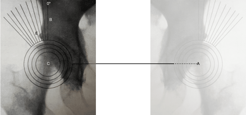
Centre-edge angle of Wiberg (1939)
Centre-edge angle of Wiberg is a radiographic measure of lateral acetabular coverage of the femoral head. Wiberg introduced the measurement in his 1939 thesis Studies on dysplastic acetabula and congenital subluxation of the hip joint as part of his investigation into acetabular dysplasia, subluxation and subsequent osteoarthritis.
Wiberg’s original construction derived its reference axis from the centres of both femoral heads; later authors modified the technique to the pelvic-horizontal/vertical construction commonly used today.
Wiberg classification of patellar morphology (1941)
In 1941 Wiberg published Roentgenographics and Anatomic Studies on the Femoropatellar Joint and proposed a classification of normal patellar morphology according to the relative sizes of the medial and lateral articular facets. Wiberg described three normal patellar types. Some later secondary sources refer to four types, but the original 1941 publication defines three.
- Type I – median ridge approximately central; medial and lateral facets roughly equal.
- Type II – ridge displaced medially; medial facet smaller than the lateral facet.
- Type III – medial facet very small, with marked dominance of the lateral facet.
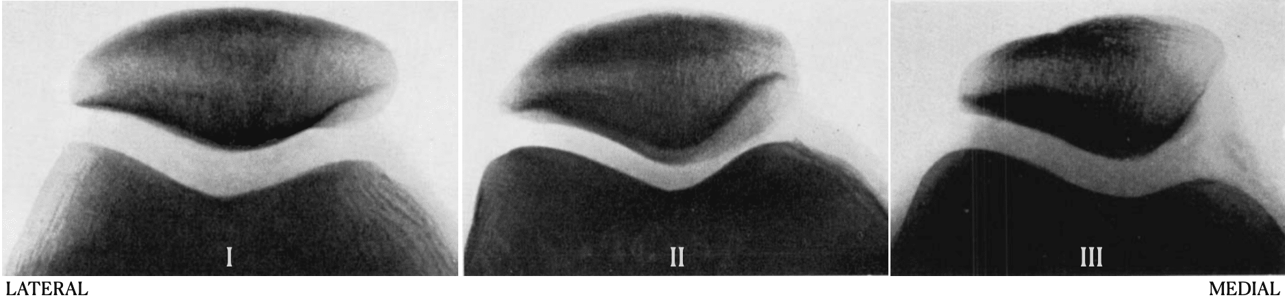
Wiberg confirmed the radiographic classification with arthrographic examination and related patellar shape to the corresponding femoral articular surface.
Key Medical Contributions
Hip dysplasia and secondary osteoarthritis: Investigated the relationship between acetabular dysplasia, congenital subluxation and later degenerative hip disease. He helped establish the concept that abnormal acetabular coverage and joint mechanics could predispose to secondary osteoarthritis.
Patellofemoral anatomy and chondromalacia: Proposed that abnormal distribution of contact and friction across the patellofemoral joint might contribute to chondromalacia patellae, particularly on the medial facet, while acknowledging that trauma and other mechanisms could also contribute.
Slipped capital femoral epiphysis: Wiberg studied epiphysiolysis capitis femoris (SCFE). He emphasised early diagnosis, and later published on wedge osteotomy for severe slips and on surgical treatment with special reference to nail fixation.
Major Publications
- Wiberg G. Studies on dysplastic acetabula and congenital subluxation of the hip. With special reference to the complication of osteoarthritis. Acta chirurgica Scandinavica 1939;83(58):5-135.
- Wiberg G. Relation between Congenital Subluxation of the Hip and Arthritis Deformans. Acta Orthopaedica Scandinavica, 1939; 10(1-4), 351–371.
- Wiberg G. Behandlung Der Epiphyseolysis Capitis Femoris Mittelst “Nagelung”. Acta Orthopaedica, 1941; 12(1-4), 179–213.
- Wiberg G. Roentgenographics and Anatomic Studies on the Femoropatellar Joint: With Special Reference to Chondromalacia Patellae. Acta Orthopaedica, 1941; 12(1-4), 319–410.
References
Biography
- Gunnar Wiberg. Acta Orthopaedica Scandinavica. 1969; 40: 411-412
- Bauer GCH. Gunnar Wiberg 1902–1988, Acta Orthopaedica Scandinavica, 1988; 59(6): 732-737
- Wiberg, Georg Gunnar Fredrik. Svenska män och kvinnor : biografisk uppslagsbok
Eponymous terms
- Severin E. Contribution to the knowledge of congenital dislocation of the hip joint: late results of closed reduction and arthrographic studies of recent cases. Acta Chir Scand. 1941;84(Suppl 63).
- Cooperman DR, Wallensten R, Stulberg SD. Acetabular dysplasia in the adult. Clin Orthop Relat Res. 1983 May;(175):79-85.
- Hanson JA, Kapron AL, Swenson KM, Maak TG, Peters CL, Aoki SK. Discrepancies in measuring acetabular coverage: revisiting the anterior and lateral center edge angles. J Hip Preserv Surg. 2015 Jun 13;2(3):280-6.
- Cao L, Sun K, Yang H, Wang H, Zeng R, Fan H. Influence of Patellar Morphology Classified by Wiberg Classification on Knee Joint Function and Patellofemoral Tracking After Total Knee Arthroplasty Without Patellar Resurfacing. J Arthroplasty. 2021 Sep;36(9):3148-3153.
- Gather KS, Sporer F, Tsagkaris C, Götze M, Gantz S, Hagmann S, Dreher T. Modified Center-Edge Angle in Children with Developmental Dysplasia of the Hip. Journal of Imaging. 2025; 11(1):3.
- Vinge R. Adult Hip Dysplasia – Prevalence and Consequence. Doctoral Thesis Department of Clinical Sciences, Lund 2025
Eponym
the person behind the name

David Yu, M.D., Ph.D. Full time anesthesiologist in the department of Perioperative medicine and Intensive Care (PMI), Karolinska University Hospital, Sweden. Dual board certified in internal medicine, and anesthesiology and critical care. Clinical interests are the critically ill patient, hemodynamic cases, perioperative echocardiography and procedural awareness, and of course all things ECG! | Squiggler |
BA MA (Oxon) MBChB (Edin) FACEM FFSEM. Emergency physician, Sir Charles Gairdner Hospital. Passion for rugby, medical history, medical education, and asynchronous learning #FOAMed evangelist. Editor-in-Chief and Webmaster of Life in the Fast lane, LITFL | On Call 4e| Eponyms | Books | Horology |