![]()
Henning Waldenström
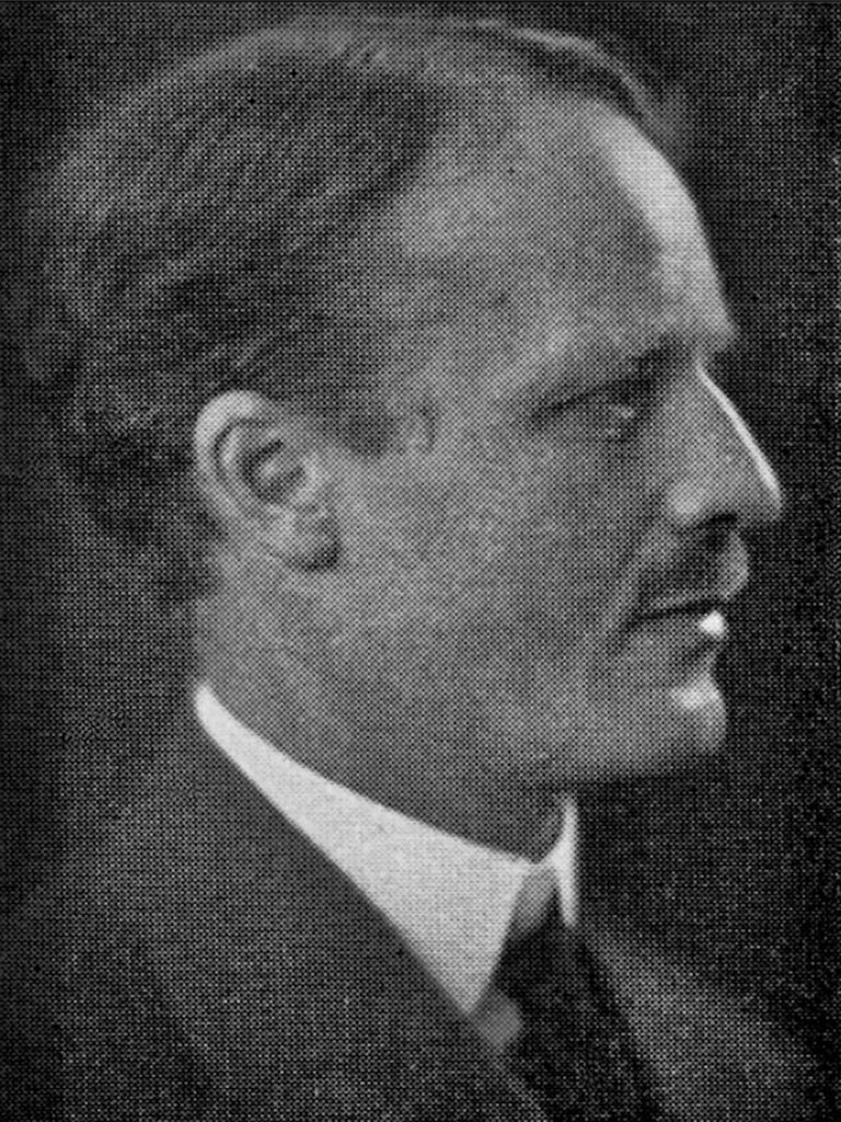
Johan Henning Waldenström (1877-1972) was a Swedish Orthopaedic surgeon.
Waldenström specialised in bone and joint tuberculosis, in particular the juvenile femoral head necrosis and epiphysiolysis capitis femoris. He also made a significant contribution to the histological examination of amyloid deposition in spleen and liver.
In 1909, Henning Waldenström described a condition of the hip-joint in 10 children under the age of 9 years which he called ‘the upper tuberculous focus in the collum‘.
1910 he provided a more detailed report of the disease in his dissertation ‘Die Tuberkulose des collum femoris im Kindersalte ihre Beziehungen zur Huftgelenkentzundung‘; expanded the number of cases to 12 and maintained a tuberculous cause.
Much to the chagrin of Waldenström, the condition later became known as Legg-Calvé-Perthes disease
Biographical Timeline
- Born on August 14, 1877 Uppsala, Sweden. Son of Physician Johan Anton Waldenström (1839-1879)
- Studied medicine in Uppsala and Stockholm; Surgical assistant at Serafimer hospital under John Wilhelm Berg (1851-1931), the father of Swedish surgery
- 1906 – Father to Jan Gösta Waldenström (1906-1996) who first described macroglobulinemia (1944)
- 1910 – Pediatric orthopedic surgeon, Stockholm
- 1929 – Founding member of Société Internationale de Chirurgie Orthopédique et de Traumatologie (SICOT)
- 1936 – Professor of orthopedic surgery Karolinska Institute, Stockholm. Specialised in surgery of the spine.
- Died January 23, 1972 in Stockholm
Medical Eponyms
Legg-Calvé-Perthes disease (LCPD); (1909, 1910)
LCPD develops as a result of proximal femoral epiphysis ischaemia of unknown aetiology; aka avascular necrosis (AVN) of the proximal femoral head.
The disease is usually insidious in onset and may occur after an injury to the hip. It is most common in male children aged 4-10 years, unilateral in 90% of cases. In cases which are bilateral, the joints are involved successively, not simultaneously.
1909 – Henning Waldenström described a condition of the hip-joint in 10 children under the age of 9 years which he called ‘the upper tuberculous focus in the collum‘. He provided a more detailed report of the disease in his dissertation ‘Die Tuberkulose des collum femoris im Kindersalte ihre Beziehungen zur Huftgelenkentzundung‘, expanded the number of cases to 12 and maintained a tuberculous cause.
Original
English
1. Fall: Knabe Erich, 14 Jahre alt. Im Alter von 8 Jahren begann der Patient zu hinken, zeitweise konnte er sich nicht auf das rechte Bein stützen… der Arzt der Schule rügte ihn wiederholt, daß er unordentlich wäre, und doch ordentlich zu gehen versuchen sollte…
weiter stellte sich bei der Untersuchung heraus, daß er hinkte, passive Bewegungen waren frei außer Abduktion un Auswärtsrotation; Trochanter aufwärts geschoben. Also das klinische Bild einer Coxa vara…
Röntgenuntersuchung (Fig 1.)…Das Collum bildet mit dem Femurschaft einen Winkel, der kleiner als normal ist. Diese Varusstellung ist… unten durch eine Krümmung des Collum bedingt, oben aber durch eine Zerstörung der oberen Kante des Collum, das dadurch eine mehr horizontale Richtung bekommen hat. Hier liegt also nicht nur eine einfache Krümmung… eine Destruktion ist auch dagewesen…
Das Caput ist in seiner Gesamtheit abgeplattet, nach oben vorgeschoben und hinaus auf den oberen Teil des Collum… so daß ein Teil der oberen Partie des Caput außerhalb der Pfanne liegt und sogar eine Eindrückung von dem oberen Rande der Pfanne erhalten kann
First case: Lad Erich, 14 years old. At the age of 8 the patient began to limp, at times he couldn’t lean on his right leg… the school doctor reprimands him repeatedly, that he is improper, and that he should try to walk properly…
Examination revealed that he limps, passive movements are unrestricted, except abduction and internal rotation; the trochanter is pushed upwards. So the clinical picture of a Coxa Vara…
X-ray examination (Fig 1.)… The femoral neck forms an angle with the shaft, which is smaller than usual. This varus position is… caused below by a deformity of the neck, above however, by a destruction of the upper edge of the neck, which has thereby acquired a more horizontal orientation. This is therefore not just a simple deformity… a destruction has also taken place…
The femoral head is flattened in its entirety, displaced upwards and outwards on the upper part of the neck… such that the upper part of the head lies partially outside the acetabulum, and even receives an impression from the superior border of the latter.
Above a fantastic and very detailed description by Waldenström of the clinical picture. Unfortunately for him, he postulated (inaccurately) that the aetiology must be infective in nature, tuberculous to be precise. In his paper he makes mention to “foci” innumerable times, which is in reference to the “focal infection theory”- a belief held by many clinicians at the turn of the 20th century, that localized foci of infection were the aetiological cause of many chronic and systemic diseases.
Original
English
…ein 10jähriger Knabe mit denselben Symptomen und gleichen Konturen auf dem Röntgenbilde wie bei dem eben beschriebenen Fall. Dieser main Fall (Nr.2) hätte also auch unter essentielle Coxa Vara gerechnet werden sollen, wenn nicht die Röntgenaufnahme (Fig 2) mir einen großen Herd- mit allen Zeichen für tuberkulösen Charakter- im oberen Teile des Collum am Caput gezeigt hätte. Der Herd schien einige Sequester zu enthalten, was…dafür sprach, daß er primär war und also die Veränderungen im Caput wahrscheinlich sekundär von Tuberkulose im Collum.
… a 10 year old lad with the same symptoms, and identical contours on x-rays, as in the previously described case. This, my second case, could also have been classified as essential coxa vara, had the X-ray not shown me a large focus- with all signs of Tuberculous character- in the upper part of the femoral head (Fig 2). The focus appears to contain several sequestrations which…speaks for it being primary in aetiology, and thus that the changes in the femoral head are secondary to Tuberculosis in the neck.

1910 – Legg, Calvé and Perthes independently reported a hip disease in children with a symptomatic picture resembling that described by Waldenström in 1909. These authors believed the process to be unrelated to tuberculosis, and the condition became known as Legg-Calvé-Perthes disease
1922 – Waldenström classification defines 4 radiographic stages of LCPD during the active phase of the disease, termed the initial stage, fragmentation stage, re-ossification stage, and residual stage, according to characteristic radiographic features.

Key Medical Attributions
Significant contribution to the histological examination of amyloid deposition in spleen and liver. Waldenström biopsied the spleens and livers of 10 patients with generalized amyloidosis and noted in all of them the gradual, occasionally complete disappearance of deposits of amyloid. In subsequent articles, he added the results of histologic studies, revealing the disappearance of amyloid which had been present before [1928;63:479-530]
Controversies
If you are looking for information on macroglobulinemia or other Waldenström eponyms – best to pop over to Henning’s son – Jan Gösta Waldenström
Major Publications
- Waldenström H. Der obere tuberkulöse Cullumherd. Zeitschrift für Orthopädische Chirurgie 1909; 24: 487–512. [Legg-Calvé-Perthes disease]
- Waldenström H. Die Tuberkulose des collum femoris im Kindersalte ihre Beziehungen zur Huftgelenkentzundung, Stockholm. 1910
- Waldenström H. The Definite Form of the Coxa Plana. Acta Radiologica, 1922; 1(4): 384-394
- Waldenström H. On coxa plana. Osteochondritis deformans coxae juvenilis. Leggs disease, maladie de Calvé, Perthes krankheit. Acta chirurgica Scandinavica. 1923; 55: 577–590.
- Waldenström H. Uber das Entstehen und Verschwinden des Amyloids beim Menschen. Klinische Wochenschrift. 1927; 47: 2235-2247
- Waldenström H. On the Formation and Disappearance of Amyloid in Man. Acta chirurgica Scandinavica. 1928; 63: 479-530
- Waldenström H. The definite form of the coxa plana. Acta Radiol 1:384–394.
- Waldenström H. The First Stages of Coxa Plana. Acta Orthopaedica Scandinavica. 1934; 5: 1-34
- Waldenström H. The First Stages of Coxa Plana. Journal of Bone and Joint Surgery. 1938; 20: 559
References
Biography
- Mostofi SB. Henning Waldenström 1877–1972. In: Who’s Who in Orthopedics. Springer, London 2005: 345
- Bibliography. Waldenström, Henning. WorldCat Identities
Eponymous terms
- Schulitz KP, Dustmann HO. Henning Waldenström (1877–1972). In: Morbus Perthes: Ätiopathogenese, Differentialdiagnose, Therapie und Prognose. Springer. 1991: 6-8
Eponym
the person behind the name

Resident medical officer in emergency medicine MB ChB (Uni. Dundee) MRCS Ed. Avid traveller, yoga teacher, polylinguist with a passion for discovering cultures.
BA MA (Oxon) MBChB (Edin) FACEM FFSEM. Emergency physician, Sir Charles Gairdner Hospital. Passion for rugby; medical history; medical education; and asynchronous learning #FOAMed evangelist. Co-founder and CTO of Life in the Fast lane | On Call: Principles and Protocol 4e| Eponyms | Books |