


Centre-edge angle of Wiberg
The centre-edge (CE) angle of Wiberg is a radiographic measure of lateral acetabular coverage of the femoral head on an AP pelvis radiograph.
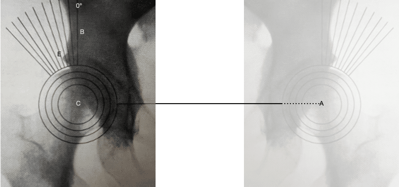
In modern practice, the angle is formed at the centre of the femoral head (C) between a vertical reference through the femoral-head centre and a second line extending from C to the lateral edge of the acetabular roof (E). A smaller angle indicates reduced lateral femoral-head coverage.
Gunnar Wiberg (1902–1988) introduced the measurement in his 1939 thesis on acetabular dysplasia, congenital hip subluxation and secondary osteoarthritis. It is now commonly termed the lateral centre-edge angle (LCEA) to distinguish it from later centre-edge measurements such as the anterior centre-edge angle.
The modern LCEA is not always constructed exactly as Wiberg described it in 1939. Both the reference axis and the definition of the lateral acetabular endpoint have changed over time..
Wiberg’s original construction
Wiberg first determined the centres of both femoral heads (A and C). The line joining those centres established the transverse pelvic reference. A perpendicular through the centre of the hip being assessed represented the longitudinal axis of the body and defined 0°.
The second limb of the CE angle extended from the femoral-head centre to point E, the lateral edge of the acetabular roof. Wiberg described E as the point where the acetabular border began to curve laterosuperiorly and where the dense shadow of the acetabular roof ended.
He regarded CE angles >25° as normal, <20° as pathological, and 20–25° as uncertain.
History of the Centre-edge angle of Wiberg
1939 – Gunnar Wiberg introduced the centre-edge angle in his thesis Studies on dysplastic acetabula and congenital subluxation of the hip. The measurement quantified the relationship between lateral acetabular coverage and femoral-head position and formed part of his investigation into dysplasia, subluxation and subsequent osteoarthritis.
1941 – Erik Severin published Contribution to the knowledge of congenital dislocation of the hip joint. He incorporated the CE angle into radiographic assessment and follow-up of congenital hip dislocation in children and extended its use into paediatric age groups.
1950 – In Congenital dislocation of the hip, William K. Massie and M. Brickett Howorth modified Wiberg’s angle for serial assessment of the developing hip. Their overlay was aligned with the horizontal plane of the pelvis, rather than deriving the transverse reference from the line joining the two femoral-head centres. A perpendicular through the centre of the femoral head then formed the vertical limb of the CE angle.
The modification was intended to permit comparison of the femoral head–acetabular relationship from childhood through adult life and across serial radiographs. This pelvic-horizontal construction is substantially closer to the form of the CE angle used today.
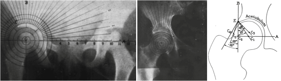
Left: measurement in a 3-year-old child. Because the epiphysis is not yet symmetrical, the overlay is centred over the midpoint of the metaphysis.
Middle: adult measurement using the same overlay, with line A aligned to the horizontal plane of the pelvis.
Right: schematic demonstrating how displacement of the femoral-head centre alters the measured CE angle.
Massie and Howorth adapted Wiberg’s method to provide a reproducible pelvic reference across growth.
1961 – Ian K. Sharp published Acetabular dysplasia: the acetabular angle. He identified several limitations of Wiberg’s CE angle as a direct measure of acetabular dysplasia. He observed that the femoral-head centre could be difficult to locate when the head was deformed. He noted that subluxation and loss of joint space altered the measurement and that displacement of the contralateral hip could tilt Wiberg’s bilateral head-centre reference. Sharp proposed an acetabular measurement independent of femoral-head position which became known as the Sharp acetabular angle.
1961 – Michel Lequesne and S. de Sèze extended Wiberg’s centre-edge concept to assessment of acetabular coverage in different planes. They introduced the false-profile view for assessment of anterior femoral-head coverage.
1963 – Lequesne published Coxométrie in which he distinguished the VCE angle on the AP pelvis (Wiberg’s CE angle referenced to the vertical and measuring lateral coverage), from the VCA angle on the false profile. This VCA angle measured anterior coverage. Lequesne noted that his VCE terminology and coxometric method dated to his thesis La coxarthrose in 1954.
1987 – Dietrich Tönnis published his textbook on Congenital Dysplasia and Dislocation of the Hip in Children and Adults. He described the CE angle using a line through the femoral-head centre parallel to the longitudinal body axis and a second line to the lateral acetabular roof. He summarised age-dependent normal values, with mean CE angles of approximately 25° at 5–8 years, 30° at 9–12 years and 35° after 13 years. He cautioned that the angle is unreliable in younger children when the centre of the incompletely ossified femoral head cannot be identified accurately.
1990 – Ogata et al. published Acetabular cover in congenital dislocation of the hip. They demonstrated that the outermost apparent bony acetabular rim and the more medial lateral end of subchondral roof condensation could diverge substantially in dysplastic hips. They proposed measuring the CE angle to the latter landmark and termed this the “refined” CE angle, finding it more closely reflected femoral-head coverage in younger children.
2010 – Shi et al. measured 2,988 hips in a Chinese population and demonstrated the strong age dependence of the CE angle. Mean values increased from 23.1° at ages 4–9 years, to 28.9° at 10–18 years, and 32.8° in adults, reinforcing that CE-angle interpretation requires consideration of age and population.
2015 – Hanson et al. published Discrepancies in measuring acetabular coverage and directly compared CE angles measured to the lateral bony edge and to the lateral sourcil. Bone-edge measurements averaged 4° greater, and 24% of hips differed by more than 5°, sometimes changing diagnostic classification. The authors revisited Wiberg’s original text and argued that his description of the dense acetabular roof shadow most likely referred to the sourcil.
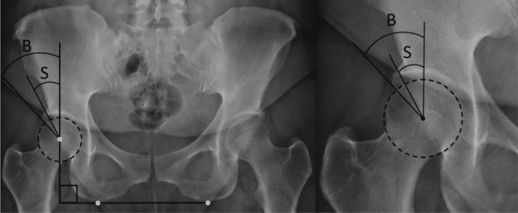
2025 – Rebecka Vinge revisited Wiberg’s original thesis in her doctoral thesis on Adult Hip Dysplasia. She concluded that his lateral endpoint corresponded to the lateral sourcil rather than the outermost bony acetabular rim. On this interpretation, Ogata’s 1990 “refined” CE angle may represent a return to Wiberg’s original landmark rather than a new modification.
2025 – Gather et al. proposed a modified CE angle for younger children using the epiphyseal joint centre and a reference perpendicular to Hilgenreiner line, intended to extend the principle of femoral-head coverage measurement into ages where the classic CE angle is limited by incomplete ossification.
Associated Persons
- Gunnar Wiberg (1902-1988)
- Dietrich Tönnis (1927-2010)
- Jørgen Reimers (1931-2008)
- Ian K. Sharp
- Michel Lequesne (1924-2022)
Alternative names
- Wiberg angle
- Centre-edge angle (CE angle)
- Lateral centre-edge angle (LCEA)
- Lateral center-edge angle
Eponymythology
One angle, two drifts
The modern lateral centre-edge angle of Wiberg is not identical to the construction Wiberg published in 1939. Both the reference axis and the lateral acetabular endpoint have drifted…
Reference axis: Wiberg derived his vertical from the line joining the centres of both femoral heads. Massie and Howorth replaced this in 1950 with a horizontal reference related to the pelvis, avoiding dependence on the position of the opposite hip. This modified construction became the standard while retaining Wiberg’s name.
Acetabular endpoint: Wiberg described point E as the lateral end of the dense acetabular roof shadow. Later literature interpreted this as either the outermost bony rim or the lateral sourcil. Ogata’s 1990 “refined” angle used the lateral roof condensation. In 2015 Hanson demonstrated that the choice of endpoint can materially alter the measured angle and clinical classification.
Re-examination of Wiberg’s original text suggests that the sourcil may have been his intended landmark. If so, the later “refined” CE angle may actually represent a return to Wiberg’s original construction.
Comparison of CE-angle values across studies requires knowing which reference axis and which lateral acetabular landmark were used
References
Historical references
- Wiberg G. Studies on dysplastic acetabula and congenital subluxation of the hip. With special reference to the complication of osteoarthritis. Acta chirurgica Scandinavica 1939;83(58):5-135.
- Severin E. Contribution to the knowledge of congenital dislocation of the hip joint: late results of closed reduction and arthrographic studies of recent cases. Acta Chir Scand. 1941;84(Suppl 63).
- Massie WK, Howarth MB. Congenital dislocation of the hip. Part I. Method of grading results. J Bone Joint Surg Am. 1950 Jul;32-A(3):519-31.
- Lequesne M. La coxarthrose. Étude clinique, anatomique et radiologique. Paris: Thèse, 1954.
- Lequesne M, Djian A. Les nouvelles incidences radiographiques pour l’étude de la hanche. Le “faux profil” du bassin. Vie Médicale. 1961;42:1629–1641.
- Sharp IK. Acetabular dysplasia: the acetabular angle. J Bone Joint Surg Br. 1961;43-B(2):268–272.
- Lequensne M, de SEZE. [False profile of the pelvis. A new radiographic incidence for the study of the hip. Its use in dysplasias and different coxopathies]. Rev Rhum Mal Osteoartic. 1961 Dec;28:643-52.
- Lequesne M. Coxométrie. Mesure des angles fondamentaux de la hanche radiographique de l’adulte par un rapporteur combiné. Rev Rhum Mal Osteoartic. 1963 Jul;30:479-85.
- Tönnis D. Normal values of the hip joint for the evaluation of X-rays in children and adults. Clin Orthop Relat Res. 1976 Sep;(119):39-47.
- Reimers J. The stability of the hip in children. A radiological study of the results of muscle surgery in cerebral palsy. Acta Orthop Scand Suppl. 1980;184:1-100.
- Tönnis D, Legal H, Graf R. Congenital Dysplasia and Dislocation of the Hip in Children and Adults. 1987
Eponymous term review
- Ogata S, Moriya H, Tsuchiya K, Akita T, Kamegaya M, Someya M. Acetabular cover in congenital dislocation of the hip. J Bone Joint Surg Br. 1990 Mar;72(2):190-6.
- Shi YY, Liu TJ, Zhao Q, Zhang LJ, Ji SJ, Wang EB. The normal centre-edge angle of Wiberg in the Chinese population: a population-based cross-sectional study. J Bone Joint Surg Br. 2010 Aug;92(8):1144-7.
- Hanson JA, Kapron AL, Swenson KM, Maak TG, Peters CL, Aoki SK. Discrepancies in measuring acetabular coverage: revisiting the anterior and lateral center edge angles. J Hip Preserv Surg. 2015 Jun 13;2(3):280-6.
- Proenca AL, Caetano AP, Bogalho L. Adult Hip Radiography: Lines and angles. ESSR 2017 / P-0209
- Radiography of the Hip: a systematic approach. ESSR 2019 / P-0091
- Bagwala J, Soni S. Evaluation of CE angle of Wiberg for the functional outcome in various hip pathologies: an observational study. Int J Res Orthop. 2019 Jan;5(1):182-188
- Gather KS, Sporer F, Tsagkaris C, Götze M, Gantz S, Hagmann S, Dreher T. Modified Center-Edge Angle in Children with Developmental Dysplasia of the Hip. Journal of Imaging. 2025; 11(1):3.
- Vinge R. Adult Hip Dysplasia – Prevalence and Consequence. Doctoral Thesis Department of Clinical Sciences, Lund 2025
- Kyle E, Cadogan M. Hip and pelvic lines and arcs. LITFL
eponymictionary
the names behind the name

MB BAO BCh Trinity College Dublin, MSc Clinical Data Analytics University of Galway. Junior doctor in Perth, doctoral researcher in MRI radiomics. Working towards radiology training.
BA MA (Oxon) MBChB (Edin) FACEM FFSEM. Emergency physician, Sir Charles Gairdner Hospital. Passion for rugby, medical history, medical education, and asynchronous learning #FOAMed evangelist. Editor-in-Chief and Webmaster of Life in the Fast lane, LITFL | On Call 4e| Eponyms | Books | Horology |