![]()
Horner Syndrome
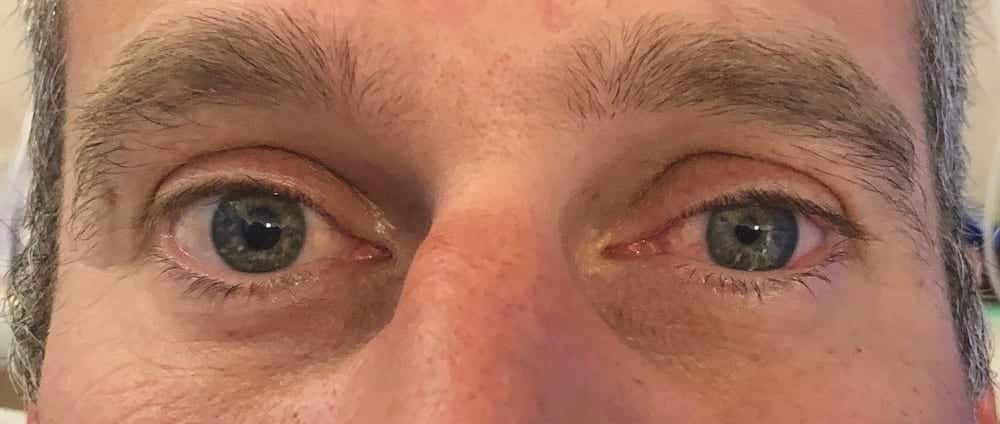
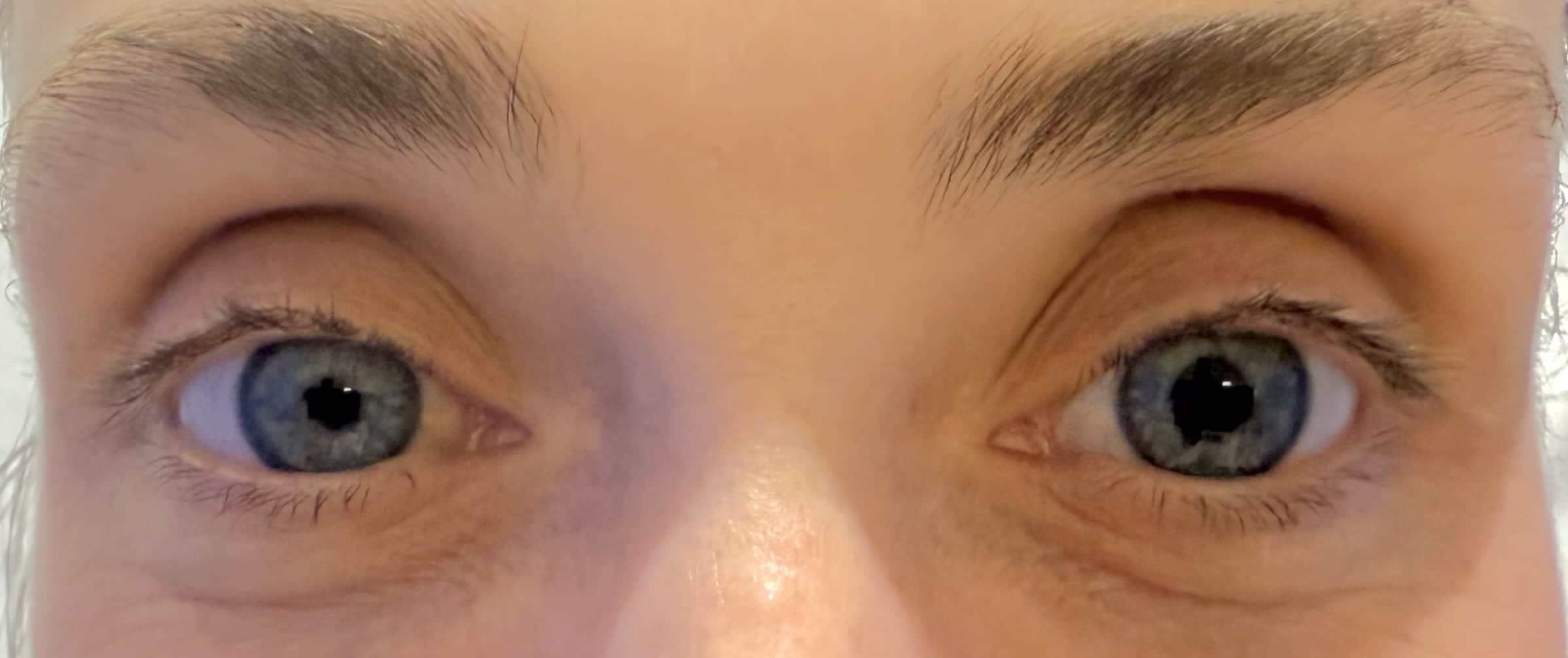
Horner syndrome, also known as oculosympathetic paresis, is a neurological disorder caused by disruption of the sympathetic pathway from the hypothalamus to the eye and face. It manifests with a classic triad of:
- Ptosis (drooping upper eyelid)
- Miosis (constricted pupil)
- Anhidrosis (reduced sweating on the affected side of the face)
Additional signs may include enophthalmos (sunken eye) and facial flushing, particularly in congenital cases or central lesions.
Aetiology
Horner syndrome results from interruption of the oculosympathetic pathway, which spans from the hypothalamus to the orbit in a three-neuron chain:
- Central (First-order neuron) lesions: Originate in the hypothalamus and descend to the spinal cord (C8–T2).
- Brainstem stroke (e.g., Wallenberg syndrome, Babinski–Nageotte syndrome, Cestan–Chenais syndrome)
- Demyelinating disease (e.g., multiple sclerosis)
- Spinal cord trauma or syringomyelia
- Pre-ganglionic (Second-order neuron) lesions: From the spinal cord to the superior cervical ganglion (through thoracic outlet and neck).
- Pancoast tumour, mediastinal mass
- Klumpke palsy (Klumpke syndrome)
- Neck trauma or iatrogenic injury (e.g., surgical dissection, epidural anaesthesia)
- Post-ganglionic (Third-order neuron) lesions: From the superior cervical ganglion via the internal carotid plexus to the eye.
- Carotid artery dissection
- Cavernous sinus lesions, cluster headaches
- Raeder paratrigeminal syndrome, Villaret syndrome
Transient or iatrogenic causes:
- Regional anaesthesia (e.g., interscalene brachial plexus block)
- Epidural analgesia
- Migraine and cluster headaches
Paediatric causes:
- Congenital birth trauma
- Neuroblastoma (thoracic or cervical)
Clinical Evaluation
History: Investigate trauma, neck pain, headache, malignancy history, recent surgery, or anaesthesia.
Examination: Careful inspection of pupil size, eyelid position, facial sweating pattern, and associated neurological deficits.
Investigations
Imaging:
- CT or MRI brain and neck to assess for central lesions or malignancy.
- CT angiography in suspected carotid artery dissection.
- Chest X-ray or CT thorax in adults with risk factors for apical lung tumour.
Pharmacological Testing:
- Apraclonidine test: Reversal of anisocoria and elevation of ptosis in the affected eye (supersensitivity of the denervated pupil).
- Cocaine test (historical): Lack of dilation in the affected pupil.
- Hydroxyamphetamine: Differentiates between pre- and post-ganglionic lesions.
Management
Primary aim: Identify and treat the underlying cause.
- In trauma or dissection: vascular consultation and anticoagulation as needed.
- In malignancy: oncology referral for staging and treatment.
- Idiopathic or transient cases: often monitored with supportive care.
Prognosis depends on the underlying pathology. Isolated idiopathic or post-procedural cases often resolve spontaneously, whereas malignancy-related or vascular causes may carry a guarded prognosis.
History of Horner Syndrome
1727 – French anatomist and surgeon François Pourfour du Petit (1664-1741) was the first to describe the ocular effects of cervical sympathetic injury in animals. In his paper Sur ce que le nerf intercostal fournit des Esprits aux yeux, he described ocular effects (miosis and ptosis) following cervical spinal transection in dogs:
Si l’Intercostal étant coupé à un Animal , il s’en ensuit des effets sensibles dans les Yeux, et qui ne puissent être rapportes à aucune autre cause…
Petit, 1727
1795 – William Cruikshank (1745–1800) Performed cervical sympathetic dissections under the direction of John Hunter, noting ocular changes and structural details of the cervical sympathetic chain, but made no clinical correlations.
1838 – British surgeon Edward Selleck Hare (1812-1838) described the case of a man who had died of a tumour on his neck. On examination he described ptosis and miosis in a patient with a cervical tumour compressing the brachial plexus. The clinical case author Hare, died on September 28, 1838, a day before the communication was published.
…a small tumour in the inferior triangular space on the left side of the neck…the pupil of the left eye became contracted; and the levator palpebrae ceased to perform its office.
Hare ES. London Medical Gazette, 1838
1839 – Scottish physiologist John Reid (1809-1849) drew attention to Hare’s case, and asked whether injury to the sympathetic chain in man might not cause constriction of the pupil as he had found it to do in animals.
…compression or section of the trunk of the sympathetic in the neck in dogs and cats is instantly followed by contraction of the pupil, the forcing of the cartilaginous membrane over the inner part of the anterior surface of the eyeball, the retraction of the eyeball deeper into the socket, and a slight approximation of the eyelids.
Reid, 1839
1852 – French physician and physiologist Claude Bernard (1813-1878) described vasomotor control by the cervical sympathetic nerve in rabbits. Bernard conducted severed the cervical sympathetic nerve and meticulously documented the ensuing physiological changes. He coined the concept of “vasomotor nerves.” and is recognised in France as co-eponym: Bernard-Horner syndrome.
After the section of the cephalic branch of the great sympathetic, it is possible to observe a contraction of the pupil of the corresponding eye, accompanied by a narrowing of the palpebral opening, a retraction of the ocular globe, and an increase of the circulation, as well as of the temperature, in all parts of the corresponding face
Bernard, 1852
1864 – American neurologist Silas Weir Mitchell (1829-1914) detailed the classic clinical triad in a 24 year old soldier with a gunshot wound to the right side of his neck:
The pupil of the right eye is very small, that of the left eye unusually large. There is a slight, but very distinct ptosis of the right eye and its outer angle appears as though it were dropped a little lower than the inner angle…his face became distinctly flushed on the right side only when walking in warm weather…a case of injury of the sympathetic nerve, probably the only one recorded.
Silas Weir Mitchell, 1862
1865 – Sir Jonathan Hutchinson (1828-1913) reported further cases with sympathetic denervation but did not claim eponym.
1869 – Swiss ophthalmologist Johann Friedrich Horner (1831-1886) reported the findings of ptosis, miosis, enophthalmos in a 40-year-old peasant woman in Über eine Form von Ptosis. He also observed increased skin temperature and dryness of the ipsilateral face.
He pharmacologically confirmed the impairment of sympathetic innervation to the eye after noting poor dilation of the affected pupil following instillation of atropine and preserved pupillary constriction to the parasympathomimetic agent calabar.
Original
English
Frau Anna Brändli, 40 Jahre alt, eine gesund aussehende Bäuerin mittlerer Grösse… bemerkte ein allmähliges herabsinken des rechten oberen Augenlides, das sehr langsam zunahm… Das obere Lid deckt die rechte Cornea bis an den oberen Pupillarrand… Die Pupille des rechten Auges ist bedutend enger als diejenige des linken… Nun erst erzählte uns die Patientin, dass sie rechterseits nie geschwitzt habe
Die Untersuchungen ergaben also Integrität des sensiblen Trigeminus, transitorische Lähmung des vasomotorischen Fasern im rechtseitigen Trigeminusgebiet;… (es) wundert sich Niemand, wenn ich diese… nie vollständige Ptosis als Lähmung des vom Sympathicus versorgten organischen Musc. palpebral super. ansehe.
Anna Brändli, aged 40, healthy looking peasant woman…noticed a slight drooping of her right upper eyelid, which increased very gradually… The upper lid covers the right cornea to the upper edge of the pupil; the lid is not loose or wrinkled but somewhat sunken into the orbit and is still capable of movement; it is neither injected nor swollen.
The pupil of the right eye is considerably more constricted than that of the left, but reacts to light; the globe has sunk inward very slightly. During the clinical discussion … the right side of her face became red and warm; while the left side remained pale and cool.
The vasomotor disturbance involves not only the trigeminal area, but also the fibres of the cervical sympathetic; this experiment with belladonna and calabar speaks for the dual control of the movements of the iris in man… we are dealing with right dilator paralysis. Ptosis a paralysis of the musculus palpebrae superioris supplied by the sympathetic nerve (H. Muller, Harling), and the appearance of the upper lid as part and parcel of the whole symptom-complex.
1873 – William Nicati (1850-1931) Horner’s student, published “La paralysie du nerf sympathique cervical“, expanding on Horner’s case and acknowledging predecessors Pourfour du Petit and Claude Bernard. Nicati’s work played a key role in cementing the eponym “Horner’s syndrome” in medical literature.
Late 1800s – Early 1900s – The term “Horner’s syndrome” gained widespread use in Germany and Switzerland. In France and Italy, the eponym “Claude Bernard-Horner syndrome” or simply “Bernard syndrome” became customary, reflecting Bernard’s earlier and more comprehensive animal model experiments.
1918 – American ophthalmologist Arnold Knapp acknowledged Horner’s contribution and noted the dual eponym usage: “These symptoms generally go under the name of Horner’s syndrome, to which the French add the name of Claude Bernard.”
1932 – Henry Pancoast (1875-1939) described the classic presentation of Horner syndrome in association with apical lung tumors, leading to the identification of the “Pancoast tumour” as a common cause of preganglionic Horner syndrome.
Mid-20th Century – Advancements in neuroanatomy and imaging refined understanding of the oculosympathetic pathway and its clinical correlations. Textbooks began routinely including Horner syndrome under neurology, ophthalmology, and thoracic pathology.
Late 20th Century – Present – The syndrome remains a clinical cornerstone for localising lesions along the three-neuron sympathetic pathway, especially in cases of stroke (e.g., Wallenberg syndrome), carotid artery dissection, and thoracic malignancy.
2000s–2020s – Ongoing refinement of diagnostic testing with pharmacological agents (e.g., apraclonidine, cocaine, hydroxyamphetamine) has improved clinical sensitivity for detecting and localizing lesions. High-resolution imaging (MRI, CTA) is now standard in evaluating new-onset Horner syndrome.
Today “Horner syndrome” is the globally accepted term, though in academic contexts, “Bernard-Horner syndrome” remains in use in Francophone countries. Historical awareness has prompted discussions about more descriptive naming conventions (e.g., “oculosympathetic paresis”), though the eponym persists due to its clinical familiarity and brevity.
Associated Persons
- François Pourfour du Petit (1664–1741) — First to describe the ocular signs experimentally.
- Edward Selleck Hare (1812-1838) — First published human clinical case.
- John Reid (1809–1849) — Experimental confirmation of sympathetic control over ocular function.
- Claude Bernard (1813-1878) — Defined the vasomotor role of the cervical sympathetic.
- John William Ogle (1824-1905) — Reviewed literature and defined the clinical triad in animals
- Silas Weir Mitchell (1829-1914) — Early full human description of the classical triad.
- Sir Jonathan Hutchinson (1828-1913) — Supplemented early human cases.
- Johann Friedrich Horner (1831-1886)— Provided detailed clinical, anatomical, and pharmacological correlation.
Alternative names
- Horner’s syndrome
- syndrome de Horner
- Bernard-Horner syndrome (France)
- Oculosympathetic paresis
- Von Passow syndrome
References
Original articles
- Petit. Sur ce que le nerf intercostal fournit des Esprits aux yeux. Histoire de l’Académie royale des sciences, 1727: 9-13
- Hare ES. Tumor involving certain nerves. London Medical Gazette 1838; 23: 16-18
- Reid J. On the Effects of Lesion of the Trunk of the Ganglionic System of Nerves in the Neck upon the Eyeball and Its Appendages. Edinb Med Surg J. 1839 Jul 1;52(140):36-43.
- Bernard C. Recherches expérimentales sur le grand sympathique et spécialement sur l’influence que le section de ce nerf exerce sur la chaleur animal. 1852
- Ogle JW. On the Influence of the Cervical Portions of the Sympathetic Nerve and Spinal Cord upon the Eye and its Appendages, illustrated by Clinical Cases, with Observations. Med Chir Trans. 1858; 41:3 97-440
- Mitchell SW, Morehouse GR, Keen WW. Chapter IV: Wounds of special nerves; wound of the sympathetic nerve. in: Gunshot Wounds and Other Injuries of Nerves. 1864: 39-44
- Horner JF. Über eine Form von Ptosis. Klinische Monatsblätter für Augenheilkunde 1869;7:193-198
- Nicati W. La paralysie du nerf sympathique cervical. Dissertation inaugurale présentée a la faculté de médecine de Zürich. Lausanne et Paris, 1873.
Eponymous review
- von Passow A. Okulare Paresen im Symptomenbilde des “Status dysraphicus”, zugleich ein Beitrag zur Ätiologie der Sympathikusparese (Horner-Syndrom und Heterochromia iridis). Münchener medizinische Wochenshrift, 1934; 74: 1243-1249.
- Kisch B. Horner’s syndrome, an American discovery. Bull Hist Med. 1951 May-Jun;25(3):284-8.
- Onuigbo WI. John Reid (1809-49) and Horner’s syndrome. Scott Med J. 1958 May;3(5):218-20.
- Durham DG. Congenital hereditary Horner’s syndrome. AMA Arch Ophthalmol. 1958 Nov;60(5):939-40.
- Miller JM. Whose eponym? The case for Edward Selleck Hare. Md Med J. 1994 Jun;43(6):498-9.
- Gao Z, Crompton JL. Horner Syndrome: A Practical Approach to Investigation and Management. Asia Pac J Ophthalmol (Phila). 2012 May-Jun;1(3):175-9.
- Walid T, Mondher BA, Mohamed Anis L, Mustapha F. A Case of Horner’s Syndrome following Ultrasound-Guided Infraclavicular Brachial Plexus Block. Case Rep Anesthesiol. 2012;2012:125346.
- Abbas A, Manjila S, Singh M, Belle V, Chandar K, Miller JP. Johann Friedrich Horner and the Repeated Discovery of Oculosympathoparesis: Whose Syndrome Is It? Neurosurgery. 2015 Sep;77(3):486-91; discussion 491.
- Prolonged Horner’s syndrome after intraoperative interscalene block 2017
- Jabua M, Gognadze T. Horner’s Syndrome Caused by Ultrasound-Guided Supraclavicular Nerve Block. Beyoglu Eye J. 2025 Mar 25;10(1):55-57.
FOAMed review and cases
- LITFL Clinical Case – Ophthalmology Befuddler 025
- CCC – Horner syndrome
eponymictionary
the names behind the name
BA MA (Oxon) MBChB (Edin) FACEM FFSEM. Emergency physician, Sir Charles Gairdner Hospital. Passion for rugby; medical history; medical education; and asynchronous learning #FOAMed evangelist. Co-founder and CTO of Life in the Fast lane | On Call: Principles and Protocol 4e| Eponyms | Books |