


Imaging of COVID-19 pneumonia: a critical care perspective
Author: Dr Greg Brogan – Peer Reviewer: A/Prof Chris Nickson
OVERVIEW
COVID-19 lung imaging is generally indicated in any COVID-19 patient with worsening respiratory status
- commonly manifests as ground glass opacities (CT chest), distributed bilaterally in bases and peripheries
- evolves rapidly and lung involvement is associated with severity; findings progress over 1-3 weeks, typically peaking at 10-14 days
- findings may be present in asymptomatic individuals or pre-symptomatic individuals and may be absent early in the course of disease
In general, the lung imaging findings of COVID-19 patients are consistent with other viral pneumonias – there is no proven specific finding for COVID-19, though there may be suggestive patterns.
CHEST RADIOGRAPHY
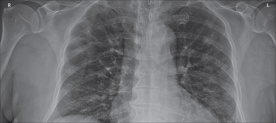
Abnormalities are seen on chest x-ray (CXR) and are more common in severe COVID-19 disease
- CXR has low sensitivity compared to RT PCR – estimates of 59-69% (Guan, WJ et. al. 2020, Wong, HYF, et al. 2020)
- Findings progress with time and appear worse at day 10-12 (Wong, HYF, et al. 2020)
- Findings may even be present before patients test positive on RT PCR (Wong, HYF, et al. 2020)
Features Include (Guan, WJ et. al. 2020 Wong, HYF, et al. 2020)
- Bilateral shadowing (72.9%) – mostly ground glass opacity (68.5%)
- Unilateral disease (25%)
- Local patchy shadowing
- Interstitial abnormalities (less common finding, <5% in some studies)
- Pleural effusions are uncommon
COMPUTED TOMOGRAPHY (CT)
Abnormalities are seen on CT in a majority of COVID-19 cases (86% (Guan, WJ et. al. 2020))
- Involved lung is proportional to severity of disease (Zhao, W et al. 2020)
Common CT features (Guan, WJ et. al. 2020 Ye, Z et al 2020 Zhao, W et al. 2020)
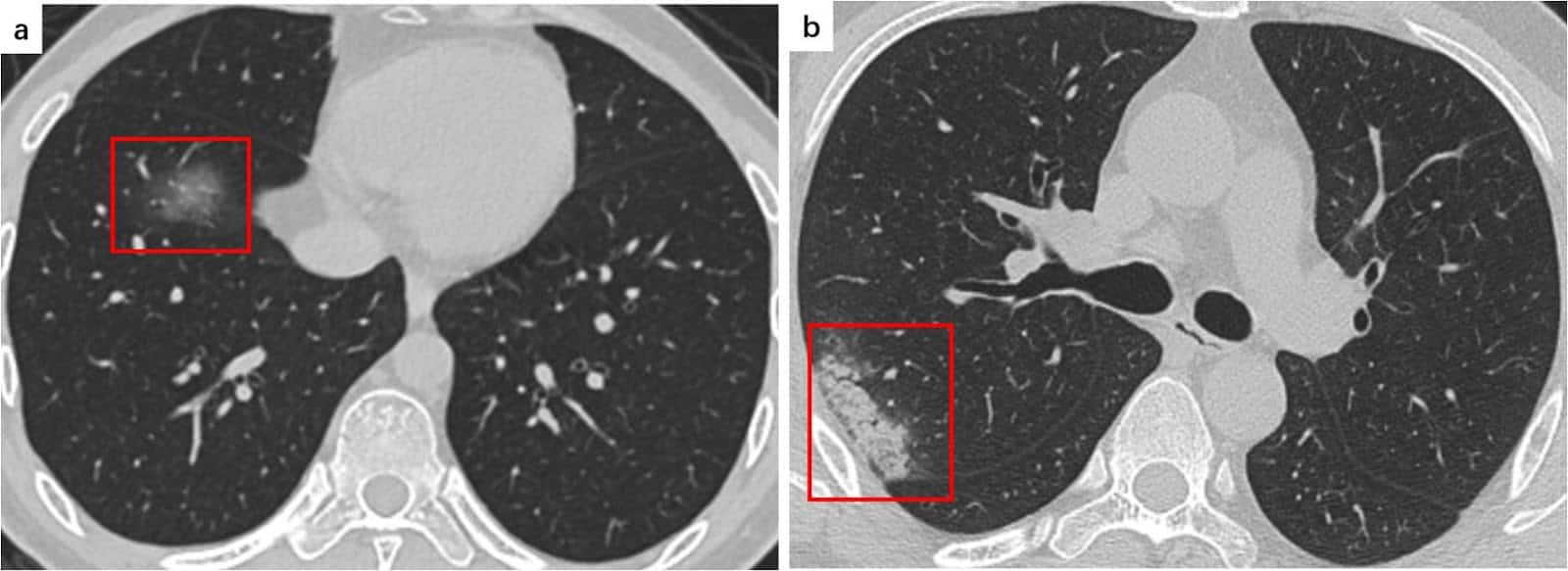
- Ground Glass Opacities (GGOs)(Ye, Z et al 2020)
- Hazy areas with increased density and no obscuring of bronchial or vascular markings (see figure 2)
- Most common manifestation – in approximately 88% of cases (Salehi S et al. 2020)
- Most often bilateral (87.5%), peripherally distributed (76%) and multilobar (78.8%) (Salehi S et al. 2020)
- Reticular Pattern (Ye, Z et al 2020)
- Multiple small linear opacities (see figure 2)
- Common, perhaps second most common pattern to GGOs
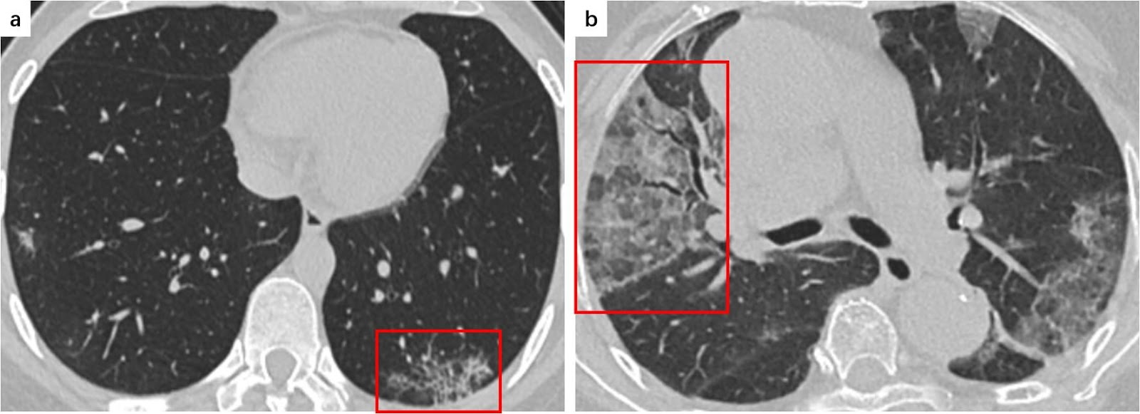
- Consolidation (Ye, Z et al 2020)
- Air spaces replaced with fluid which obscures margins of vessels and airways (see figure 3)
- Sign of progression of disease
- Multifocal, patchy or segmental
- Often subpleural or along bronchovascular bundles
- Crazy Paving Pattern (Ye, Z et al 2020)
- Resembles irregular paving stones from thickened interlobular septa/intralobular lines superimposed onto GGO (see figure 3)
- Seen in 5-36%
Uncommon CT manifestations include (Salehi S et al. 2020, Rodriguez JCL. et al 2020)
- Pleural effusion
- Pericardial effusion
- Lymphadenopathy
- Cavitation
- CT Halo sign
- Pneumothorax
Super-infection may be suggested by pleural effusion, extensive tiny lung nodules, tree-in-bud and lymphadenopathy (Rodriguez JCL. et al 2020)
When followed over time, a pattern to stages of disease emerges (Li, M et al 2020)
- Early Phase – Moderate clinical manifestations with lesions limited to single or multiple areas
- Progressive phase – Lesions progress in extent and density with the accumulation of cellular exudate in alveoli
- Severe Phase – Pulmonary lesions reach a peak at around 14 days with dense bilateral infiltration and a large amount of cellular exudate
- Dissipative Phase – after 14 days, gradual absorption of lesions occurs (may occur earlier if disease course shorter)
Role of CT in diagnosis:
- A single study of 1014 patients in Wuhan suggested high sensitivity (97%), but low specificity (25%) to detect disease the during outbreak (Ai, T et al. 2020)
- A small study of CT-radiologists were able differentiate COVID from non-COVID viral pneumonias (Bai, HX 2020), however they had a wide range of sensitivity (67-93%) and specificity (7-100%)
- Advantage of faster availability than PCR tests, though non-specific and risks of transport remain
- Changes can even be seen in asymptomatic patients (Shi, H et al. 2020)
- However, findings are time dependent (Rodriguez JCL. et al 2020) – Approximately half may have normal study 2 days after symptoms onset.
Lung Ultrasonography (US)
The role of lung ultrasonography is still being defined, but no specific COVID-19 findings have been confirmed (Soldati G et al. 2020, Vetrugno, L et al 2020)
- Only sparse case series and case reports currently available
Uses
- May help reduce the amount of CT scans and X-rays required (Vetrugno et al 2020)
- May provide a useful alternative in a climate where transport capabilities, staffing and imaging availabilities are stressed (Smith et al 2020)
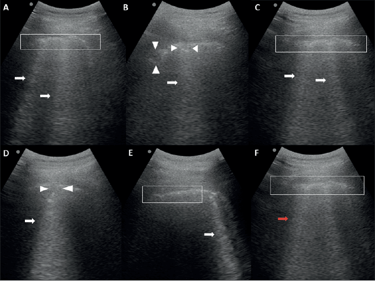
Cases exhibit presence of viral pneumonia with features including (Buonsenso, D et al. 2020):
- Irregular pleural line
- B-lines (may be irregular and even confluent)
- Patchy pattern with bilateral sparing
- Areas of white lung
- Subpleural consolidations
Risks
- It is important to consider infection control and prevention of transmission via contact with ultrasound machine
References
LITFL
- LITFL Coronavirus resources
- Coronavirus 2019 (COVID-19) (overview from the Critical Care Compendium)
- Lung Ultrasound in COVID-19
Journal articles
- Ai T, Yang Z, Hou H, et al. Correlation of Chest CT and RT-PCR Testing in Coronavirus Disease 2019 (COVID-19) in China: A Report of 1014 Cases [published online ahead of print, 2020 Feb 26]. Radiology. 2020;200642. doi:10.1148/radiol.2020200642
- Bai HX, Hsieh B, Xiong Z, et al. Performance of radiologists in differentiating COVID-19 from viral pneumonia on chest CT [published online ahead of print, 2020 Mar 10]. Radiology. 2020;200823. doi:10.1148/radiol.2020200823
- Buonsenso D, Piano A, Raffaelli F, Bonadia N, de Gaetano Donati K, Franceschi F. Point-of-Care Lung Ultrasound findings in novel coronavirus disease-19 pnemoniae: a case report and potential applications during COVID-19 outbreak. Eur Rev Med Pharmacol Sci. 2020;24(5):2776–2780. doi:10.26355/eurrev_202003_20549
- Guan WJ, Ni ZY, Hu Y, et al. Clinical Characteristics of Coronavirus Disease 2019 in China [published online ahead of print, 2020 Feb 28]. N Engl J Med. 2020;10.1056/NEJMoa2002032. doi:10.1056/NEJMoa2002032
- Li M, Lei P, Zeng B, et al. Coronavirus Disease (COVID-19): Spectrum of CT Findings and Temporal Progression of the Disease [published online ahead of print, 2020 Mar 20]. Acad Radiol. 2020;S1076-6332(20)30144-6. doi:10.1016/j.acra.2020.03.003
- Rodrigues JCL, Hare SS, Edey A, et al. An update on COVID-19 for the radiologist – A British society of Thoracic Imaging statement. Clin Radiol. 2020;75(5):323–325. doi:10.1016/j.crad.2020.03.003
- Rodriguez-Morales AJ, Cardona-Ospina JA, Gutiérrez-Ocampo E, et al. Clinical, laboratory and imaging features of COVID-19: A systematic review and meta-analysis [published online ahead of print, 2020 Mar 13]. Travel Med Infect Dis. 2020;101623. doi:10.1016/j.tmaid.2020.101623
- Salehi S, Abedi A, Balakrishnan S, Gholamrezanezhad A. Coronavirus Disease 2019 (COVID-19): A Systematic Review of Imaging Findings in 919 Patients [published online ahead of print, 2020 Mar 14]. AJR Am J Roentgenol. 2020;1–7. doi:10.2214/AJR.20.23034
- Shi H, Han X, Jiang N, et al. Radiological findings from 81 patients with COVID-19 pneumonia in Wuhan, China: a descriptive study. Lancet Infect Dis. 2020;20(4):425–434. doi:10.1016/S1473-3099(20)30086-4
- Silverstein WK, Stroud L, Cleghorn GE, Leis JA. First imported case of 2019 novel coronavirus in Canada, presenting as mild pneumonia [published correction appears in Lancet. 2020 Feb 29;395(10225):e41]. Lancet. 2020;395(10225):734. doi:10.1016/S0140-6736(20)30370-6
- Smith MJ, Hayward SA, Innes SM, Miller A. Point-of-care lung ultrasound in patients with COVID-19 – a narrative review [published online ahead of print, 2020 Apr 10]. Anaesthesia. 2020;10.1111/anae.15082. doi:10.1111/anae.15082
- Soldati G, Smargiassi A, Inchingolo R, et al. Is there a role for lung ultrasound during the COVID-19 pandemic? [published online ahead of print, 2020 Mar 20]. J Ultrasound Med. 2020;10.1002/jum.15284. doi:10.1002/jum.15284
- Vetrugno L, BoveT, Orso D, et al. Our Italian experience using lung ultrasound for identification, grading and serial follow-up of severity of lung involvement for management of patients with COVID-19 [published online ahead of print, 2020 Apr 1]. Echocardiography. 2020;10.1111/echo.14664. doi:10.1111/echo.14664
- Wong HYF, Lam HYS, Fong AH, et al. Frequency and Distribution of Chest Radiographic Findings in COVID-19 Positive Patients [published online ahead of print, 2019 Mar 27]. Radiology. 2019;201160. doi:10.1148/radiol.2020201160
- Ye Z, Zhang Y, Wang Y, Huang Z, Song B. Chest CT manifestations of new coronavirus disease 2019 (COVID-19): a pictorial review [published online ahead of print, 2020 Mar 19]. Eur Radiol. 2020;10.1007/s00330-020-06801-0. doi:10.1007/s00330-020-06801-0
- Zhao W, Zhong Z, Xie X, Yu Q, Liu J. Relation Between Chest CT Findings and Clinical Conditions of Coronavirus Disease (COVID-19) Pneumonia: A Multicenter Study [published online ahead of print, 2020 Mar 3]. AJR Am J Roentgenol. 2020;1–6. doi:10.2214/AJR.20.22976
FOAM and web resources
- Radiopaedia – COVID-19
- Intensive and The Alfred ICU – COVID-19 resources
SARS-CoV-2
novel coronavirus of COVID-19

Chris is an Intensivist and ECMO specialist at The Alfred ICU, where he is Deputy Director (Education). He is a Clinical Adjunct Associate Professor at Monash University, the Lead for the Clinician Educator Incubator programme, and a CICM First Part Examiner. He is an internationally recognised Clinician Educator with a passion for helping clinicians learn and for improving the clinical performance of individuals and collectives. He was one of the founders of the FOAM movement (Free Open-Access Medical education) has been recognised for his contributions to education with awards from ANZICS, ANZAHPE, and ACEM.
His one great achievement is being the father of three amazing children.
On Bluesky, he is @precordialthump.bsky.social and on the site that Elon has screwed up, he is @precordialthump. | INTENSIVE | RAGE | Resuscitology | SMACC