![]()
Lid cracked open
aka Ophthalmology Befuddler 034
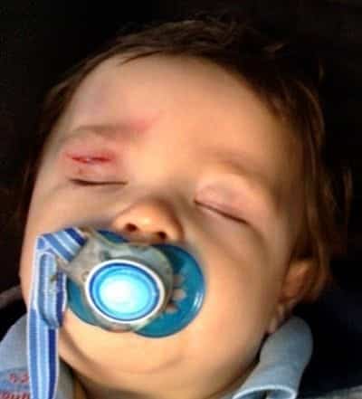
A 3 year-old boy is brought to the emergency department by his father after coming second best in a ‘head versus chair’ collision.
Questions
Q1. What is shown?
Answer and interpretation
Right Upper Eyelid laceration
Q2. What are aspects of history and examination should be included in the assessment of this injury?
Answer and interpretation
Assume a lid injury is a penetrating eye injury until proven otherwise.
History:
- mechanism, e.g. bite, foreign body, etc
- symptoms, e.g. pain, tearing, altered vision
Examination:
- visual acuity
- assess superficial structures looking for conjunctival penetration or laceration. Ideally use a slit lamp, or simply a magnifying glass.
- rule out serious eye injury before wound closure — this may require detailed fundoscopic examination depending on the history and mechanism.
- explore the wound fully to assess depth and rule out foreign bodies — lid lacerations may appear deceptively superficial.
- Damage to the nasolacrimal drainage system should be suspected if a laceration is present nasal to the upper or lower eyelid punctum. Punctal dilation and irrigation of the canalicular system may be required.
Anatomy of the lacrimul punctum (RootAtlas)
Q3. Describe the management of this injury.
Answer and interpretation
Superficial lacerations can be managed in the ED:
- univeral precautions, antisepsis, irrigation, local anesthesia (e.g. 2% lignocaine with adrenaline), debridement and removal of foreign bodies, closure with 6/0 non-absorbable sutures.
- avoid deep sutures and never suture the orbital septum, which will cause eyelid tethering.
- tetanus prophylaxis
- Antibiotics are not usually indicated — consider in contaminated wounds or following bites.
Is there more to the injury than just a lid laceration?
- Consider the need for C-spine clearance and CT head in the case of head trauma.
- Orbital XR or CT may be necessary to rule out foreign body or orbital fracture.
Q4. Which eyelid wounds require ophthalmological referral?
Answer and interpretation
Refer to an ophthalmologist if any of the following are present:
- laceration involving the lid margin
- possible damage to the nasolacrimal duct system (i.e. punctum, canaliculus, common duct, or lacrimal sac) — a laceration that is nasal to either the upper or lower eyelid punctum.
- full thickness lid laceration
- extensive tissue loss or distortion of anatomy
- medial canthal tendon avulsion (suspect when there is displacement, excessive rounding, or abnormal laxity of the medial canthus)
- involvement of the levator aponeurosis of the upper eyelid (producing ptosis) or the superior rectus muscle
- visible orbital fat in an eyelid laceration, indicating penetration of the orbital septum. Such patients require CT imaging and careful assessment of levator and extraocular muscle function.
- associated ocular trauma requiring surgery (e.g. ruptured globe, intraorbital foreign body)
References
- Ehlers JP, Shah CP, Fenton GL, Hoskins EN. The Wills Eye Manual: Office and Emergency Room Diagnosis and Treatment of Eye Disease Lippincott Williams & Wilkins
- NSW Statewide Opthalmology Service. Eye Emergency Manual — An illustrated Guide. [Free PDF]
OPHTHALMOLOGY BEFUDDLER
Chris is an Intensivist and ECMO specialist at The Alfred ICU, where he is Deputy Director (Education). He is a Clinical Adjunct Associate Professor at Monash University, the Lead for the Clinician Educator Incubator programme, and a CICM First Part Examiner.
He is an internationally recognised Clinician Educator with a passion for helping clinicians learn and for improving the clinical performance of individuals and collectives. He was one of the founders of the FOAM movement (Free Open-Access Medical education) has been recognised for his contributions to education with awards from ANZICS, ANZAHPE, and ACEM.
His one great achievement is being the father of three amazing children.
On Bluesky, he is @precordialthump.bsky.social and on the site that Elon has screwed up, he is @precordialthump.
| INTENSIVE | RAGE | Resuscitology | SMACC