
![]()
Neuro 101: Brainstem
Anatomy of the Brainstem
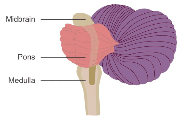
The brainstem is organised into three regions: the medulla, pons, and midbrain. It also contains the cranial nerve nuclei.
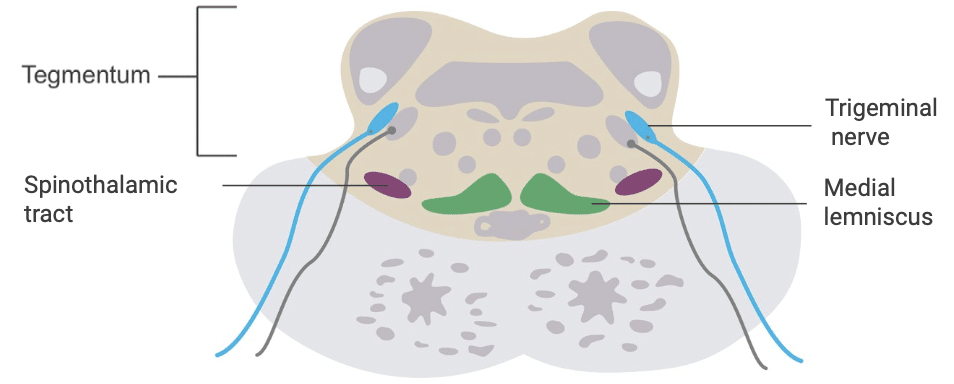
The Medulla
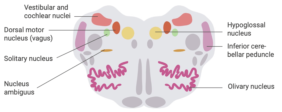
Lateral Medullary Syndrome (Wallenberg Syndrome)
Typically caused by stroke involving the posterior inferior cerebellar artery (PICA) or its branches.
Lesion involves
- Inferior cerebellar peduncle
- Vestibular nuclei
- Fibres or nuclei of cranial nerves IX and X
- Spinal nucleus and tract of cranial nerve V
- Spinothalamic tract
- Sympathetic pathways
Symptoms
- Contralateral loss of pain and temperature in body
- Ipsilateral loss of pain and temperature in face
- Dysphagia
- Dysphonia
- Decreased gag reflex
- Vertigo
- Nystagmus
- Ipsilateral ataxia
- Diplopia
- Ipsilateral Horner’s syndrome
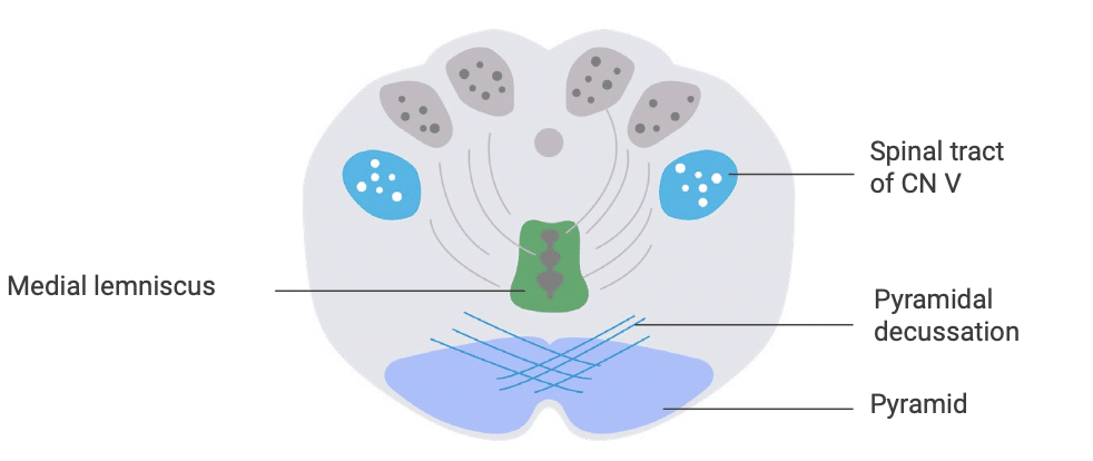
Medial Medullary Syndrome
Caused by occlusion of the anterior spinal artery or vertebral artery.
Lesion involves
- Medial lemniscus
- Hypoglossal nerve (CN XII)
- Pyramid (corticospinal tract)
Symptoms
- Contralateral body paralysis
- Ipsilateral tongue weakness (tongue deviates away from weakness)
- Contralateral hemianaesthesia of body (sparing the face)
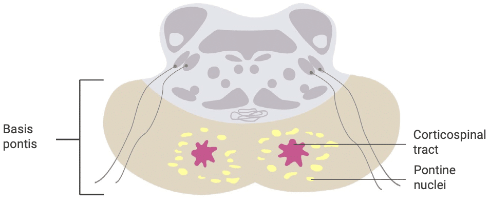
The Pons
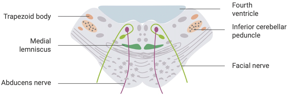
Pontine Syndromes
Usually due to small vessel lacunar strokes affecting perforating branches of the basilar artery. Lesions can occur at various levels along the long axis of the pons.
Lesions may involve
- Corticospinal tract
- Cranial nerves V, VI, or VII
Symptoms
- Contralateral hemiplegia
- Contralateral hemianaesthesia (with larger lesions)
Locked-In Syndrome
Caused by basilar artery occlusion affecting the basis pontis.
Lesion involves
- Corticospinal tracts
- Corticobulbar tracts
Symptoms
- Impaired speech
- Loss of facial movement
- Quadriplegia
- Patient is awake and aware (eye movements may be preserved)
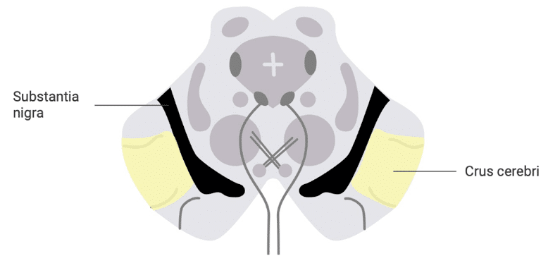
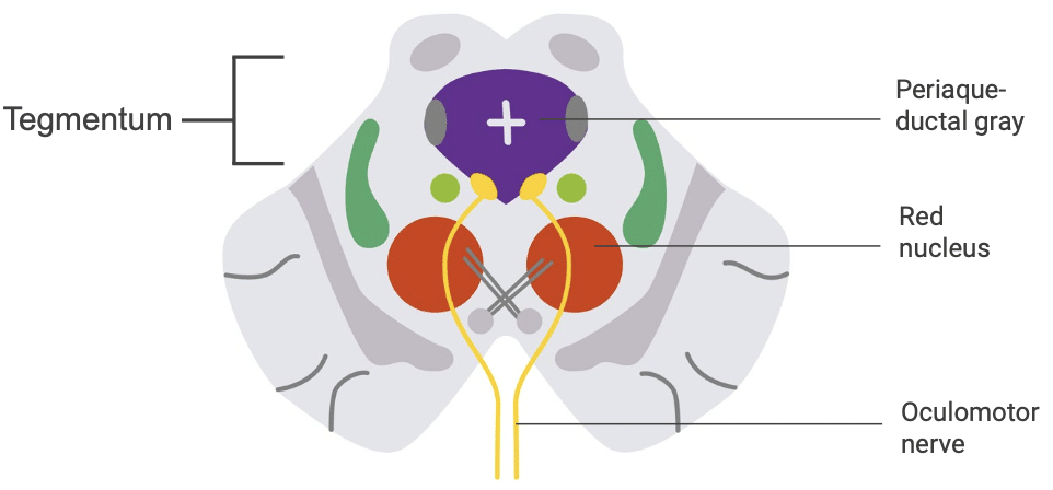
The Midbrain
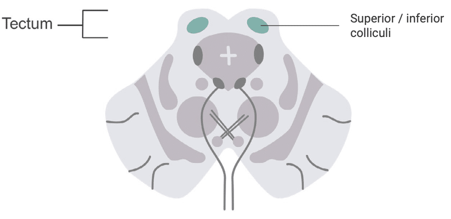
Parinaud Syndrome (Dorsal Midbrain Syndrome)
Often due to compression of the tectum (e.g., by a pineal tumour).
Lesion affects
- Inferior and/or superior colliculi
Symptoms
- Paralysis of vertical gaze
- Convergence-retraction nystagmus
- Downbeat nystagmus
- Mid-position pupil dilation (with light reactivity loss)
Benedikt Syndrome
Caused by vascular, inflammatory, or mass lesions involving the midbrain tegmentum.
Lesion affects
- Oculomotor nucleus (CN III)
- Red nucleus
Symptoms
- Ipsilateral oculomotor palsy
- Contralateral ataxia
- Intention tremor
Weber Syndrome
Often results from midbrain strokes or tumours.
Lesion affects
- Cerebral peduncle
- Oculomotor nerve (CN III)
Symptoms
- Contralateral hemiplegia
- Ipsilateral oculomotor palsy
Clinical Neurology Essentials
- Coni R. Neuro 101: Neurological Examination. LITFL
- Coni R. Neuro 101: Cerebral Hemispheres. LITFL
- Coni R. Neuro 101: Cerebellum and Basal Ganglia. LITFL
- Coni R. Neuro 101: Brainstem. LITFL
- Coni R. Neuro 101: Cranial Nerves. LITFL
- Coni R. Neuro 101: Spinal Cord. LITFL
- Coni R. Neuro 101: Peripheral Nervous System. LITFL
References
Further reading
- Brazis PW, Masdeu JC, Biller J. Localization in Clinical Neurology. 8e 2021
- Campbell WW. DeJong’s The Neurologic Examination. 8e 2019
- Fuller G. Neurological Examination Made Easy. 6e 2019
- Kiernan J. Barr’s The Human Nervous System: An Anatomical Viewpoint. 10e 2015
- O’Brien M. Aids to the Examination of the Peripheral Nervous System. 6e 2023
- Patten JP. Neurological Differential Diagnosis. 2e 1996
- Waxman SG. Correlative Neuroanatomy. 23e 1996
Publications
- Aydogdu I, Ertekin C, Tarlaci S, Turman B, Kiylioglu N, Secil Y. Dysphagia in lateral medullary infarction (Wallenberg’s syndrome): an acute disconnection syndrome in premotor neurons related to swallowing activity? Stroke. 2001 Sep;32(9):2081-7
- Boller F. Strokes and behavior: disorders of higher cortical functions following cerebral disease. Disorders of language and related function. Stroke. 1981 Jul-Aug;12(4):532-4.
- Carrera E, Michel P, Bogousslavsky J. Anteromedian, central, and posterolateral infarcts of the thalamus: three variant types. Stroke. 2004 Dec;35(12):2826-31.
- Datar S, Rabinstein AA. Cerebellar infarction. Neurol Clin. 2014 Nov;32(4):979-91
Neurology Library

Robert Coni, DO, EdS, FAAN. Vascular neurologist and neurohospitalist and Neurology Subspecialty Coordinator at the Grand Strand Medical Center in South Carolina. Former neuroscience curriculum coordinator at St. Luke’s / Temple Medical School and fellow of the American Academy of Neurology. In my spare time, I like to play guitar and go fly fishing. | Medmastery | Linkedin |