![]()

LITFL Update 045
Update 045. Sending you FOAMed content from around the globe. Latest trends and best articles so that you stay top of your field.

![]()
Update 045. Sending you FOAMed content from around the globe. Latest trends and best articles so that you stay top of your field.

A 60-year-old woman presents with immediate onset of sore throat after accidentally ingesting a foreign body.
A 35-year-old women presents 4 weeks postpartum with a day of RUQ pain and fevers. She has mildly elevated LFTs.
A 35-year-old man presents with 48 hours of sore throat, husky voice, fever and difficulty breathing when lying flat.

Nebulised GTN in the ED. Case study and review of dose, administration and indication for GTN nebuliser administration
Egerton Y. Davis IV, Head of UCEM's Demographically Impartial Public & Social Health Improvement Taskforce, made an important announcement today. Davis said that his team have been allocated the task of saving Britain's National Health Service.
A 32-year-old female presents with chills and painful right sided facial swelling.
She is normally well. Her symptoms have been present for the last 48 hours.
June 2024 Adult Abdominal imaging cases and interpretation with Kylee Brooks, Parker Hambright, Alexis Holland, William Lorenz, Brent Mathews and Kyle Cunningham
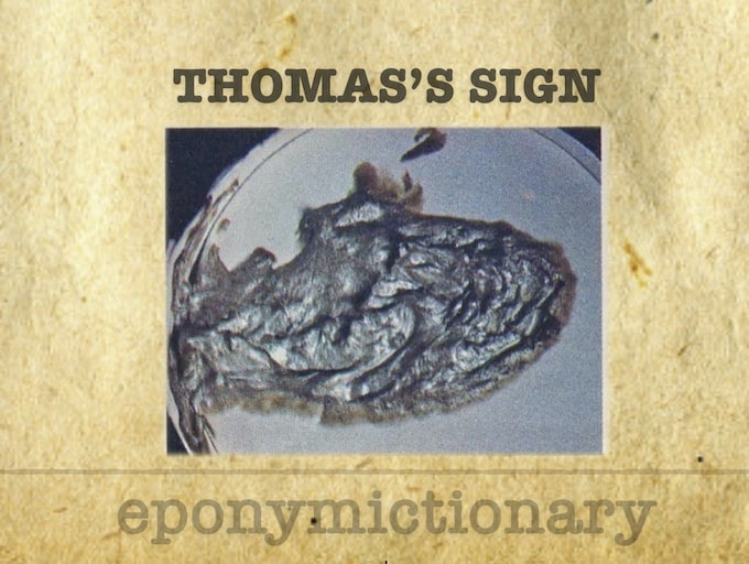
Thomas's sign: Silver, aluminium or metallic coloured stools. Caused by the combination of cholestatic (acholic) pale stools secondary to CBD obstruction and the black-tar colour malaena.
A 78-year-old lady from a nursing home is brought into ED by ambulance with acute upper abdominal pain, vomiting, and abdominal distension.
A 42-year-old lady presents with three weeks of headache. She has a past history of recurrent unprovoked pulmonary embolus
A 50-year-old unrestrained male passenger involved in a high speed MVA is brought in by the pre-hospital team to your emergency department after a 60-minute retrieval time.