![]()
Shepherd fracture
Description
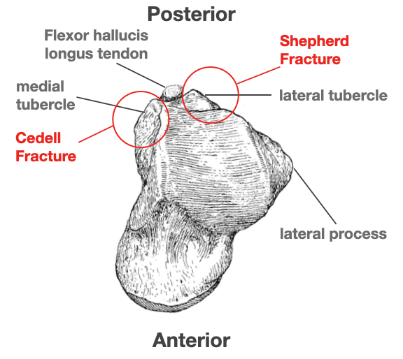
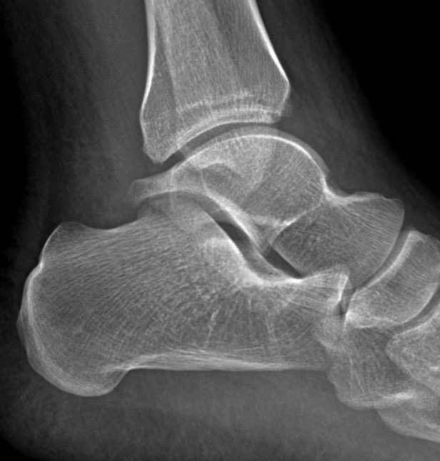
Sherpherd fracture: Posterior talar process fracture with injury to the lateral tubercle caused by inversion or extreme equinus. Otherwise known as fracture of the lateral tubercle of the posterior process of the talus.
The posterior talofibular ligament attaches to the lateral tubercle and the tendon of flexor hallucis longus runs between these the medial and lateral tubercles.
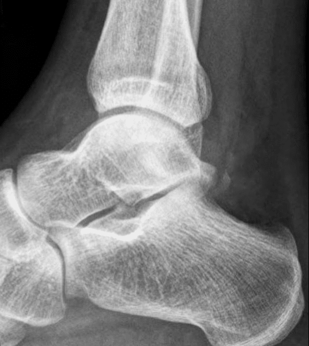
Shepherd fracture may be mistaken for an os trigonum (secondary ossification center accessory ossicle) which lies posterior to the lateral tubercle and which is a normal finding. The os trigonum is usually round/oval with smooth well corticated edges as opposed to the sharply marginated non-corticated irregular outline of a fracture
Fracture is usually seen on lateral XR, however sensitivity only 78%. In cases of significant trauma and suspicion CT or MRI should be considered.
Complications include chronic pain, arthrosis, and occasionally avascular necrosis.
Treatment is generally with immobilisation, although rarely delayed excision of the fragments may be necessary
Note: **Cedell fracture – posterior talar process fracture with injury to the medial tubercle caused by forced dorsiflexion and pronation. Shepherd fractures are more common than Cedell fractures.
History of the Shepherd fracture
1882 – Described by Francis John Shepherd (1851-1929), initially as a single dissection and later as a series of three such fractures.
Last year I exhibited to this Society (Medico-Chirurgical Society of Montreal) a specimen of fracture of the astragalus (talus) found in a subject in the dissecting-room. Since then I have examined the condition of the astragalus in every subject dissected, and have been fortunate enough to obtain two more examples of the same fracture.
The fractured portion is the little process of bone external to the groove for the tendon of the flexor longs hallucis muscle: This process is on the posterior border of the astragalus, and overhangs the os calcis. To it is attached the posterior fasciculus of the external lateral ligamnet of the ankle-joint; called sometimes the posterior peroneo-tarsal ligament. Judging from the appearance of the fracture, it would seem that the process of bone is torn off by the ligament being put on the stretch in some twist of the foot.
May not this fracture account for some cases of sprained ankles which are so slow to recover, and which occasionally leave permanent lameness, or at any rate weakness? In such cases as I have described, it is probable that any motion of the foot (as flexion and twisting out) which puts the posterior peroneo-tarsal ligament on the stretch would be painful.
Shepherd 1882
2004 – Dan-Henrik Boack and Sebastian Manegold described a modified classification system to be applied to fractures of either the lateral or the posterior processes.
Type 1: small chip or avulsion fracture (< 0.5cm)
- 1a: small (extra-articular) fragment of the lateral process of the talus;
- 1b: small fragment of the isolated medial tubercle of the posterior process;
- 1c: small (intra-articular) fragment of the lateral process of the talus.
Type 2: intermediate fragment (0.5 – 1.0cm) with some displacement
- 2a: extends into the subtalar joint but not to the talofibular joint;
- 2b: isolated fracture of the entire lateral tubercle of posterior process.
Type 3: large fracture fragment (> 1cm) with associated damage to both the ankle and the subtalar joints
- 3a: single large fragment of the lateral process extending from the talofibular articular surface to the posterior facet of the subtalar joint;
- 3b: comminuted fracture of the entire lateral process;
- 3c: fracture of the entire posterior process of the talus.
Type 4: severe form of fracture of either of the processes and associated instability or dislocation of the subtalar joint.
Stieda Process
The Stieda process is an elongated lateral tubercle of the posterior process of the talus. It is often considered an anatomical variant. A fracture through the Stieda process is termed a Stieda process fracture. Named after German anatomist Christian Hermann Ludwig Stieda (1837–1918) who first described the anatomical variant in 1869 in the paper Ueber secundäre Fusswurzelknochen.
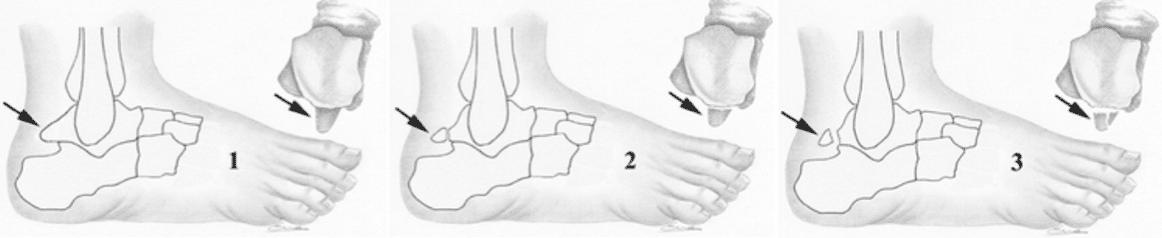
1. Stieda process; 2. os trigonum; 3. Shepherd fracture AJR 2003
Associated Persons
- Francis John Shepherd (1851-1929)
- Carl-Axel Cedell (1932-2021)
- Christian Hermann Ludwig Stieda (1837–1918)
References
Original articles
- Stieda L. Ueber secundäre Fusswurzelknochen. Archiv für Anatomie, Physiologie und Wissenschaftliche Medicin, 1869: 108-111
- Shepherd FJ. A Hitherto Undescribed Fracture of the Astragalus (talus). J Anat Physiol. 1882; 17(Pt 1): 79–81.
Review articles
- Ludwig Stieda (1837–1918). Nature, 1937; 140(3550): 840.
- Hodge JC. Clinics in diagnostic imaging (42). Shepherd’s fracture. Singapore Med J. 1999 Oct;40(10):666-8
- Haddad FS, Bartlett M, Singh D. The sequelae of posterior talar fractures. Injury. 2000 Mar;31(2):107-11
- Cerezal L et al. MR imaging of ankle impingement syndromes. AJR Am J Roentgenol. 2003 Aug;181(2):551-9
- Boack DH, Manegold S. Peripheral talar fractures. Injury. 2004 Sep;35 Suppl 2:SB23-35..
- Ahmad R, Ahmed AMY. Fracture of the posterior process of the talus: an unusual injury. Emerg Med J. 2007 Dec; 24(12): 867.
- Early JS. Talus fracture management. Foot Ankle Clin. 2008; 13(4): 635-57
- Kou JX, Fortin PT. Commonly missed peritalar injuries. J Am Acad Orthop Surg. 2009; 17(12): 775-86.
- Summers NJ, Murdoch MM. Fractures of the talus: a comprehensive review. Clin Podiatr Med Surg. 2012; 29(2): 187-203, vii
- Dale JD, Ha AS, Chew FS. Update on talar fracture patterns: a large level I trauma center study. AJR Am J Roentgenol. 2013 Nov;201(5):1087-92.
- Blanchette MA, Grenier JM. Fracture of the lateral tubercle of the posterior talar process caused by a rock-climbing fall: a case report. J Can Chiropr Assoc. 2014 Sep;58(3):286-90.
- Harvey J. Stieda process. Radiopaedia
- McKennedy C. Shepherd fracture. Eponym A Day. Instagram
- Eponymythology: Eponymous ankle and talus injuries. LITFL
[cite]
eponymictionary
the names behind the name
BA MA (Oxon) MBChB (Edin) FACEM FFSEM. Emergency physician, Sir Charles Gairdner Hospital. Passion for rugby; medical history; medical education; and asynchronous learning #FOAMed evangelist. Co-founder and CTO of Life in the Fast lane | On Call: Principles and Protocol 4e| Eponyms | Books |