


Sixth Cranial Nerve Lesions
Cranial nerve VI is also known as the Abducens nerve.
It is a purely somatic motor nerve.
Isolated lesions are uncommon, but do occur.
Multiple sclerosis is one prominent cause.
When found in association with other cranial nerve lesions, a space-occupying lesion is more likely
Anatomy
Course of the Abducens Nerve
- Originates in the abducens nucleus within the pons.
- Exits the ventral brainstem in the posterior fossa at the junction of the pons and medulla, medial to the facial nerve, which itself lies medial to the vestibulocochlear nerve.
- Passes forward over the petrous temporal bone, into the middle cranial fossa, lateral to the sella turcica, within the cavernous sinus:
- Lies lateral to the internal carotid artery
- Lies medial to the ophthalmic nerve
- Leaves the cavernous sinus and enters the orbit through the superior orbital fissure and common tendinous ring.
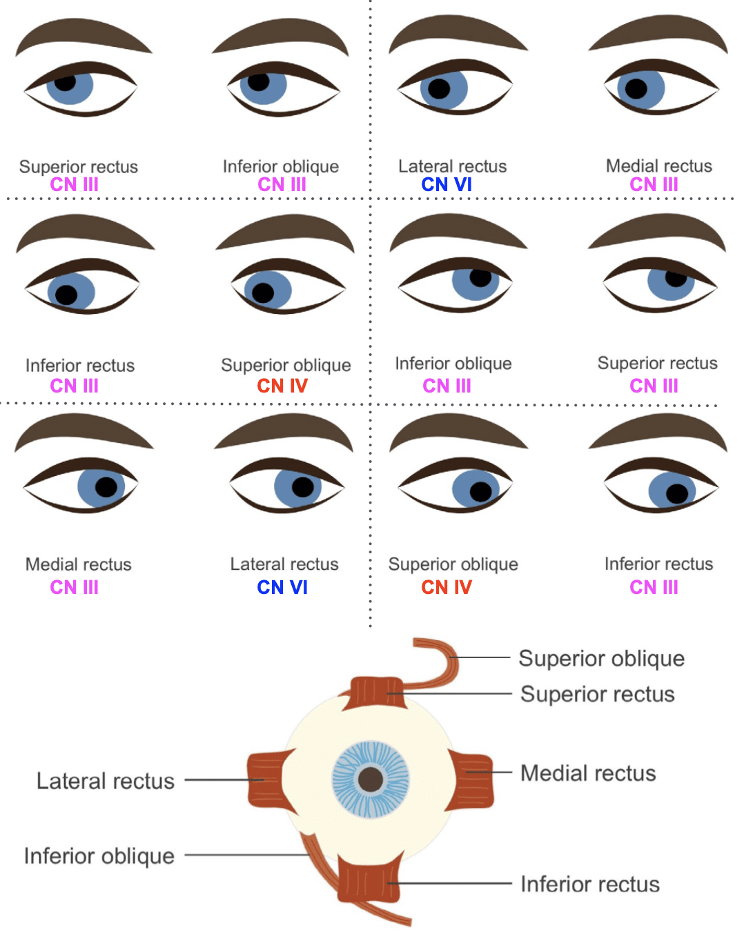
- Runs along the medial aspect of the lateral rectus muscle, which it supplies.
Abducens Nerve Innervations
| Function | Structure Innervated |
|---|---|
| Motor | Lateral rectus muscle |
Pathology
Causes of a sixth cranial nerve lesion include:
- Demyelinating disease
- Multiple sclerosis
- Vascular disease
- Brainstem microvascular strokes
- Space-occupying lesions
- Tumours
- Aneurysms
- Abscesses
- Raised intracranial pressure
- Cerebral oedema
- Intracerebral haemorrhage (ICH)
- Subarachnoid haemorrhage (SAH)
- Venoms
- Snake bite
- Thiamine deficiency
- Wernicke’s encephalopathy (manifestation of ophthalmoplegia)
- Trauma
- Especially involving the petrous temporal bone, where the abducens nerve crosses
- Mononeuritis
- Diabetes
- Toxins
- Microvascular disease
- Paraneoplastic disease
- Connective tissue disease
- Infectious disease (HIV, Lyme disease [US], syphilis)
- Idiopathic
- No clear cause found in some cases
- Rare causes
- Cavernous sinus thrombosis
- Usually in combination with lesions of other cranial nerves within the cavernous sinus
- Cavernous sinus thrombosis
- Congenital causes
- Congenital absence of the sixth nerve (e.g. Duane syndrome)
Clinical Assessment
Important Points of History
- Presenting problem usually diplopia.
- Patients may also present with a head-turned attitude in an attempt to maintain binocular vision.
Important Points of Examination
- Strabismus
- May be an obvious medially directed squint of the affected eye.
- Eye movement testing
- Failure of lateral movement of the affected eye.
- Test both eyes together, and if abnormality found, each eye separately.
- Double vision
- Signs are maximal when looking to the affected side.
- Images are horizontal and parallel.
- Outermost image (from affected eye) disappears on covering that eye.
- Outermost image usually more blurred.
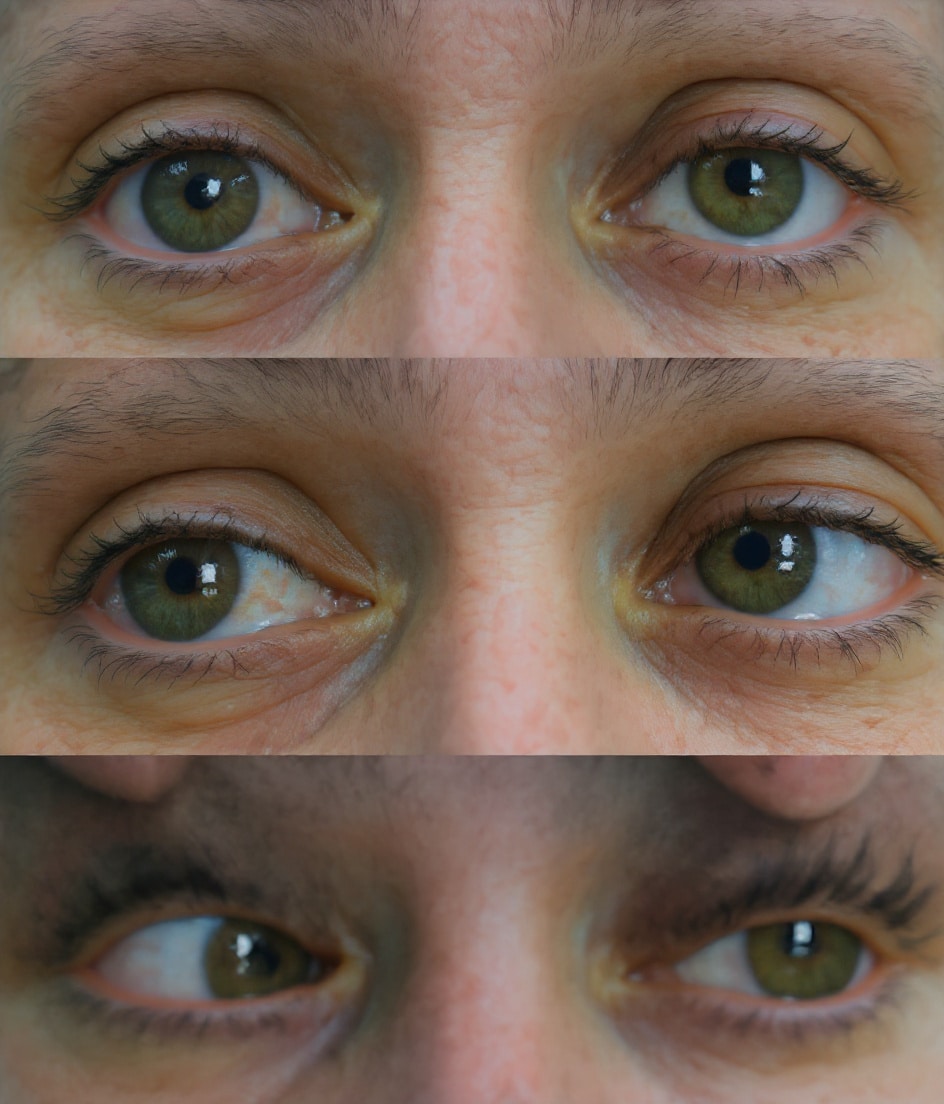
Above: Central gaze (primary position), looking straight ahead
Middle: Gaze to the right, no restriction
Below: Gaze to the left, the side of the lesion. Note failure of the left eye to fully abduct.
This woman presented to the ED with a sudden onset of severe headache and vomiting. CT angiogram scan revealed a SAH due to a ruptured left vertebral artery aneurysm.
Investigations
When Clinical Diagnosis Is Clear
- None may be necessary (e.g. in snake envenomation).
Otherwise Consider:
Blood Tests
- FBC
- CRP
- ESR
- U&Es / glucose
CT Scan / CT Angiogram
- Good screening test for intracranial mass lesions.
- CT angiogram for suspected aneurysmal disease.
MRI
- Best imaging investigation for the sixth cranial nerve.
- Especially useful for:
- Intracranial / intraorbital space-occupying lesions (tumours, abscesses, aneurysms)
- Multiple sclerosis
Management
- Management depends on the underlying cause.
Diplopia
- Patients should be warned not to perform high-risk activities (e.g. driving).
- Use of an eye patch may relieve debilitating diplopia.
Disposition
- Disposition depends largely on cause:
- Mass lesions or bleeds → Urgent referral to Neurosurgery.
- Isolated lesions in otherwise well patients → Referral to Neurology and/or Ophthalmology.
Appendix 1

Appendix 2
References
Publications
- Brazis PW, Masdeu JC, Biller J. Localization in Clinical Neurology. 8e 2021
- Fuller G. Neurological Examination Made Easy. 6e 2019
- O’Brien M. Aids to the Examination of the Peripheral Nervous System. 6e 2023
FOAMed
- Coni R. Neuro 101: Cranial Nerves. LITFL
- Nickson C. The Brainstem Rules of Four. LITFL
- Ercleve T. The rule of 4 of the brainstem. LITFL
- Nickson C. Third Cranial Nerve Lesions. LITFL
- Nickson C. Cranial nerve lesions DDx. LITFL
Fellowship Notes

MBBS FACEM DDU (Emergency) CCPU. Emergency Physician in Melbourne, Australia. Co-Ultrasound Lead for Emergency Medicine at The Alfred Hospital. Special interests in diagnostic and procedural ultrasound, medical education, and ECG interpretation | ECG Library |

Educator, magister, munus exemplar, dicata in agro subitis medicina et discrimine cura | FFS |