![]()
Sunao Tawara
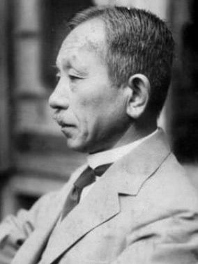
Sunao Tawara 田原 淳, (1873 – 1952) was a Japanese pathologist and teacher.
Born initially as Sunao Nakashima, Tawara would take the surname of his physician uncle after being adopted by him at the age of 15, and go on to be considered one of the fathers of modern cardiac electrophysiology.
A renowned teacher during his time, Tawara would be present for the founding of the new Kyūshū University where he would go on to lecture for 25 years and be instrumental in making their School of Medicine one of the most productive institutions for medical research in Japan.
Tawara’s contributions would greatly advance the understanding of the heart’s conduction system and become a reference for future research for years to come. It is speculated by some that his contributions were Nobel Prize-worthy, however, the political climate during Tawara’s time saw him ineligible for such prestige.
Tawara is eponymous with the Tawara node. A street in Kyūshū is also named after him to honor his achievements.
Biography
- Born July 5, 1873 in Aki, Ōita Prefecture, Kyūshū
- 1901 – Graduated in medicine from the University of Tokyo
- 1901-1902 – Studied dermatology then internal medicine at the Tokyo University Hospital
- 1902-1903 – Practiced dermatology in his father’s clinic in the Ōita Prefecture
- 1903-1906 – Research pathologist in Ludwig Aschoff’s laboratory at Philipps University of Marburg
- 1906 – Published Das Reizleitungssystem des Säugetierherzens
- 1906-1908 – Associate Professor of Pathology at the Fukuoka Medical School, Kyoto National University
- 1908 – Doctorate of medical science from the University of Tokyo
- 1908-1933 – Professor of Pathology at the University of Kyūshū (newly established in 1908 with Fukuoka Medical School being its core)
- 1914 – Awarded the Imperial Prize of the Japan Academy and given a special medal by the Emperor for his work on the heart’s conduction system
- Died January 19, 1952 in Fukuoka, Kyushu
Medical Eponyms
Tawara node (1906)
The atrioventricular node [AV node; Node of Tawara; Aschoff-Tawara node]
Tawara’s 1906 monograph, ‘Das Reizleitungssystem des Säugetierherzens‘ [The Conduction System of the Mammalian Heart] established the link between the bundle of His and the Purkinje fibers by discovering the left and the right bundle branch, the interposed components between them, and by identifying the Purkinje fibers as the terminal ramifications of these components. Tawara added another component, the atrioventricular node, and defined these components as a system – the conduction system of the heart.
Tawara explained the conduction system as a closed system, like that of an anatomic tree rooted in the atrial septum, with the stem and the main branches located in the ventricular septum. The peripheral branches extend to the papillary muscles and the parietal walls, and that the smallest ramifications of the Purkinje fibers ultimately spread, as the terminal ramifications of the conduction system, to all the parts of the ventricles.
The AV node is sometimes referred to as the Aschoff-Tawara node. However, although Tawara’s work on the conduction system was carried out in Aschoff’s laboratory and under his guidance, Aschoff did not claim to be a co-author of the monograph, only providing the foreword to Tawara’s work.
Key Medical Contributions
Tawara made significant contributions to the knowledge of the cardia conducting system. These findings had been overlooked for the best part of a century before the work of Koso Suma brought many of the findings to light. Suma summarises Tawara’s contribution to the conduction system as:
- Identification of the atrioventricular node with a reticular formation at the sites where the atrioventricular conduction system originates
- Precise description of the courses of the right and the left bundle
- Identification of the false tendons as parts of the conduction system
- Identification of the Purkinje fibers as the terminal ramifications of the conduction system
- Precise description of the histology of each part of the conduction system
- Precise estimation of the conduction velocity of excitation in the conduction system and of the excitatory process and the mode of contraction of the ventricles.
Tawara produced detailed photographic and illustrated images to demonstrate the conduction system. Below is a macroscopic image of the left ventricle of the human heart. The anterior wall of the left ventricle was cut from just below the aortic valve toward the cardiac apex at the line between the anterior and the posterior papillary muscles and opened toward the right and the left. The entire course of the left bundle branch and its terminal ramifications are illustrated.

Stimulations for further advancements
Tawara’s work had a significant impact on the progress of cardiology.
Tawara’s discovery of the atrioventricular node stimulated Keith and Flack to search for such a peculiar tissue in another region of the heart, as it was believed at that time that the heart beat was initiated in the musculature surrounding the terminal part of the superior vena cava. In 1907, Arthur Keith (1866-1955) and Martin Flack (1882-1934) reported the existence of the sino-atrial node in the vertebrate heart.
In 1903, Willem Einthoven (1860 – 1927) published a paper reporting the recording of the ECG with a string galvanometer developed by himself. This attracted little attention from the medical profession. However, following the publication of the paper, “More about the electrocardiogram,” Einthoven was besieged by visitors and received correspondence from individuals all over Europe who wanted to see or learn about the new instrument. In this publication, Einthoven referred to the contributions of Sunao Tawara 田原 淳, (1873 – 1952) as the theoretical basis for interpreting electrocardiogram.
Original
English
Nach den Untersuchungen Tawara’s besteht das atrioventrikulare Verbindüngsbfindel aus einem System vonMuskelfasern, die einen sich baumförmig verzweigenden Strang bilden…Völlig in glbereinstimmung mit diesem anatomischen Bau ist die Fortpfianzung der Kontraktionswelle im Herzen, wie dieselbe aus tier Form des Elektrokardiogrammes abgeleitet werden muss.
According to Tawara’s study, the atrioventricular connecting bundle consists of a system of muscle fibers which form a tree-like ramified cord…The mode of propagation of the contraction wave in the heart, as it must be deduced from the form of the electrocardiogram, is in complete accordance with this anatomical structure.
Major Publications
- Tawara S. Das Reizleitungssystem des Säugetierherzens: Eine anatomisch-pathlogische Studie über das Atrioventrikularbündel und die Purkinjeschen Fäden. Jena, Verlag von Gustav Fischer, 1906. [Suma K. Conduction System Of The Mammalian Heart, The: An Anatomico-histological Study Of The Atrioventricular Bundle And The Purkinje Fibers. [English translation] 2000]
- Aschoff L, Tawara S. Die heutige Lehre von den pathologisch-anatomischen Grundlagen der Herzschwäche; kritische Bemerkungen auf Grund eigener Untersuchungen. 1906.
- Tawara S. Anatomisch-Histologische Nachprüfung der Schnittführung an der von Prof. H.E. Hering übersandten Hundeherzen. Arch ges Physiol Mensch Thier 1906; 111:300–302
- Tawara S. Über die sogenannten abnormen Sehnenfäden des Herzens. Ziegl Beitr Path Anat 1906; 39:563–584.
References
Biography
- Suma K. Sunao Tawara: a father of modern cardiology. Pacing Clin Electrophysiol. 2001;24(1):88-96.
- Loukas M, Linganna S, Chiba A, Tubbs RS. Sunao Tawara, a cardiac pathophysiologist. Clin Anat. 2008;21(1):2-4.
- Akiyama, Toshio. Sunao Tawara: Discoverer of the atrioventricular conduction system of the heart. Cardiology journal. 2010;17:428-34.
- Bibliography. Tawara, Sunao 1873-1952. World Cat Identities
Eponymous terms
- Fye WB. The origin of the heart beat: a tale of frogs, jellyfish, and turtles. Circulation. 1987;76(3):493-500.
- Ehrlich W. The discovery of the cardiac conduction system: the testimony of the authors. Perspect Biol Med. 1992;35(4):487-498.
- Suma K. Tawara S. Conduction System Of The Mammalian Heart, The: An Anatomico-histological Study Of The Atrioventricular Bundle And The Purkinje Fibers. [English translation] 2000
Eponym
the person behind the name
Lewis is an RMO at Royal Perth Hospital. He is currently interested in critical care medicine.
BA MA (Oxon) MBChB (Edin) FACEM FFSEM. Emergency physician, Sir Charles Gairdner Hospital. Passion for rugby; medical history; medical education; and asynchronous learning #FOAMed evangelist. Co-founder and CTO of Life in the Fast lane | On Call: Principles and Protocol 4e| Eponyms | Books |