
![]()
Wilhem His Jr
Wilhelm His Jr. (1863–1934) was a Swiss cardiologist and anatomist.
Born the son of Dr. Wilhelm His Sr., His Jr. would follow his father’s legacy and go on to become a well-renowned figure who contributed greatly to the fields of embryological, histological, and cardiovascular electrophysiology research.
A well-rounded man, His Jr. also mastered German and French at a young age and was a proficient violinist and painter. He was described by one of his students as “a master of his profession, a great physician, investigator, and a well-cultured gentleman”,
His Jr. is eponymous with the Bundle of His and the Werner-His disease.
Biography
- Born December 29, 1863 in Basel, Switzerland. Third of six children of Dr. and Mrs. Wilhelm His Sr (1831-1904)
- 1889 – MD, University of Leipzig; Assistant to Heinrich Curschmann (1846-1910) in the Leipzig Clinic
- 1893 – Described the Bundle of His in his 35-page article on the activity of the embryonic heart
- 1895 – Professor extraordinary at the University of Leipzig
- 1901 – Chief physician at the Friedrich City Hospital, Dresden
- 1902 – Associate professor of internal medicine at Basel/
- 1907 – Director of the Charité medical clinic; Chair of internal medicine in Berlin
- 1914-1918 – Consulting internist for the German army, going on missions to Turkey, Asia Minor, the Western theater, and Russia
- 1916 – Described Trench fever whilst on a mission in Russia
- 1918 – Dean of the medical faculty in Berlin
- 1928 – Rector of the University of Berlin
- 1932 – Retired from all work duties due to emphysema
- Died November 10, 1934
Medical Eponyms
Bundle of His (1893)
The atrioventricular bundle (bundle of His) arises in the specialised tissue of the atrioventricular node (AV node, Tawara node), and continues in the interventricular septum as a single bundle, the crus commune. It divides into two trunks that pass respectively to the right and left ventricles, fine branches passing to all parts of the ventricles. The bundle of His conducts impulses from the atria to the Purkinje fibres of the ventricles, and initiates ventricular contractions.
In 1893, His Jr. reported the atrioventricular bundle. He described the bundle on one page of his 36 page dissertation:
Original
English
Nach längerem Nachforschen ist es mir jedoch gelungen, ein Muskelbündel zu finden, welches Vorhof- und Kammerscheidewand untereinander verbindet, und welches bisher der Beobachtung dadurch sich entzogen hat, dass es, bei geringem Umfange, nur dann in ganzer Ausdehnung sichtbar wird, wenn die Scheidewände genau der Länge nach getrotfen sind. Sowohl auf derartigen Schnitten, als auch in Schnittserien konnte ich den Verlauf des Bündels erkennen, und habe dasselbe bisher nachgewiesen bei einer ausgewachsenen Maus, einem neugeborenen Hunde, zwei neugeborenen und einem erwachsenen (ca. 30jährigen) Menschen (Fig. 4). Das Bündel entspringt von der Hinterwand des rechten Vorhofs, nahe der Vorhofsscheidewand, in der Atrioventricularfurche, legt sich der oberen Kante des Kammerscheidewandmuskels unter mehrfachem Faseraustausch an, zieht auf demselben nach vorn, bis es, nahe der Aorta, sich in einen rechten und einen linken Schenkel gabelt, welch letzterer in der Basis des Aortenzipfels der Mitralis endigt.
After extensive investigation I was able to find a muscle bundle which connects the atrial and ventricular septal walls, and which apparently has not been observed before, because it is only visible in its entire distribution when the septal walls are cut exactly in the horizontal direction. I was able to recognise the course of this bundle on such sections, and have proved its presence in an adult mouse, a new born-dog, two new-born infants and one adult around 30 years of age (Fig. 4). The bundle arises from the posterior wall of the ventricle near the atrial septum in the atrioventricular groove, it joins the upper edge of the ventricular septum and ramifies, coursing on the septum anteriorly until it branches near the aorta into a right and left branch, the latter terminating in the base of the aortic cusp of the mitral valve.
His 1893: 23 [Translation Willus FA. 1941]
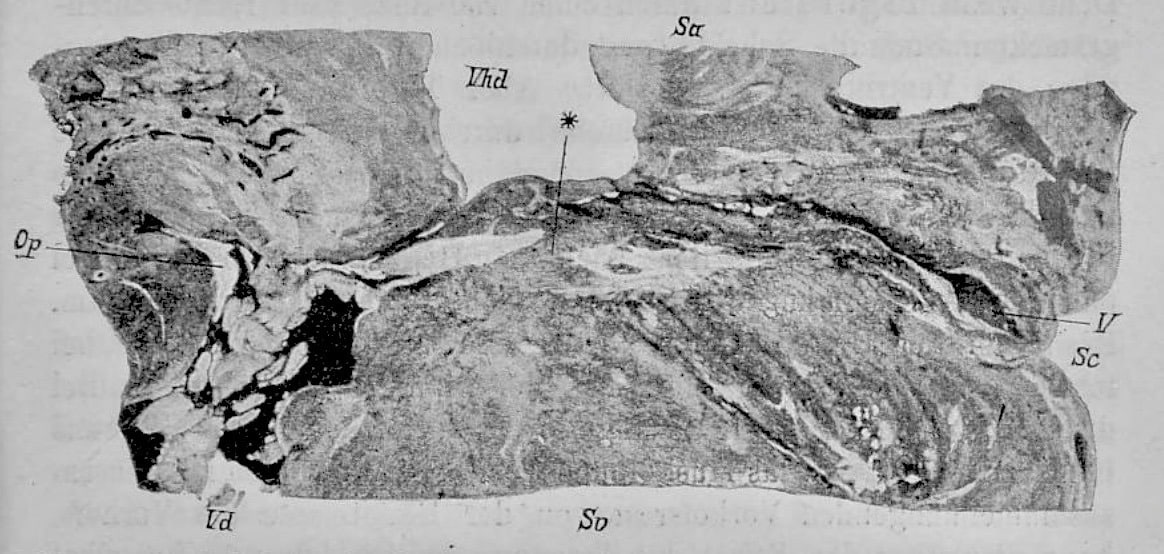
Fig. 4. Sagittal section through the atrioventricular border of the heart of a human newborn. Magnification 7.5. The ventricular septum is complete, the atrial septum cut lengthways. Vhd right atrium. Vd right ventricle. Sv Ventricular septum Sa Atrial septum. Sc coronary groove. V Coronary vein. Op Ostium pulmonale. * transition bundle.
Original
English
Ob dieses Bündel wirklich die Erregung vom Vorhof zum Ventrikel leitet, kann ich nicht mit Sicherheit angeben, da ich bisher Durchtrennungsversuche an demselben nicht angestellt habe. Jedenfalls ist dessen Vorhandensein ein Grund gegen die Meinung derjenigen, welche mit dem Mangel musculösen Zusammenhanges zwischen Vorhof und Kammer die Nothwendigkeit nervöser Leitung zu beweisen suchen.
I cannot state with certainty whether this bundle actually conducts the impulses from the atrium to the ventricle, as I did not perform any experiments dealing with the severing of the bundle. Its presence, in all events, is contrary to the opinion of those, who, in the absence of such a muscular connection between the atrium and the ventricle, attempt to prove the necessary presence of a nerve conduction
Werner-His disease (1916)
Trench fever [** aka five-day fever; quintan fever; Wolhynia fever; His disease; Meuse fever; shin bone fever] is a self-limiting Rickettsial infection which caused outbreaks affecting over 1 million soldiers in Europe during World War I. The infecting organism is Bartonella quintana (formerly Rochalimaea quintana; Rickettsia quintana), an aerobic, Gram-negative rod-shaped bacillus. The primary vector is the human body louse.
Described independently in the 1916 edition of Berliner Klinische Wochenschrift by Heinrich Werner (1874-1946) and Wilhelm His Jr
- Werner H. Über eine besondere Erkrankung, die er als Fünftagefieber bezeichnet. Berliner klinische Wochenschrift, 1916; 53(1): 204
- His Jr W. Über eine neue periodische Fiebererkrankung (Febris Wolhynica). Berliner klinische Wochenschrift, 1916; 53(1): 322-323
Key Medical Attributions
Coining the term ‘heart block’
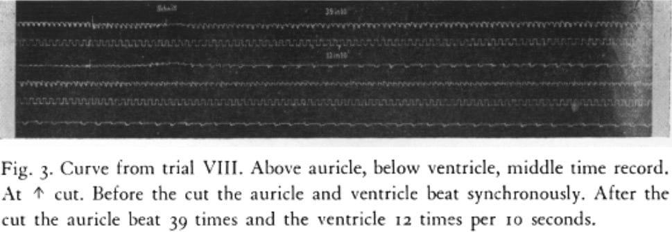
In 1893, whilst experimenting on rabbit hearts, His Jr. confirmed his theory that conduction from the atria to the ventricles occurred through the AV bundle, and that damaging this bundle resulted in an asynchronous contraction of the two chambers. His Jr. decided to wait instead of immediately publishing his findings, as he wished to show this to also be the cause of the Stokes-Adams attacks. He would be unfortunate, however, in the fact that he would only see one case of Stokes-Adams, and that one case would be unavailable for autopsy. His Jr. eventually published his findings in 1899, coining the term ‘heart block’ in the process.
Controversies
While His Jr. was not known to be an active antisemite or Nazi sympathizer, the same cannot be said with respect to his views of the mentally ill. In his official rector’s address in 1928, entitled Über die natürliche ungleichheit des menschen, His emphasized the evils of contemporary culture which he believed could be eliminated by eugenic measures.
Major Publications
- His Jr W. Die Thätigkeit des embryonalen Herzens und deren Bedeutung für die Lehre von der Herzbewegung beim Erwachsenen. Arbeiten aus der medicinischen Klinik zu Leipzig. Herausgegeben von H. Curschmann. 1893; 14 – 49. [The activity of the embryonic human heart and its significance for understanding of the heart movement in the adult Translated Bast TH, Gardner WD Journal of the History of Medicine and Allied Sciences, 1949; 4(3): 289–318.] [Bundle of His]
- His Jr W. Ein Fall von Adams-Stokes’scher Krankheit mit ungleichzeitigem Schlagen der Vorhöfe und Herzkammern (Herzblock). Dtsch Arch Klin Med 1899; 64, 316-331 [Heart Block]
- His Jr W. Antritlsrede, gehalten zum Beginn der Klinik. Berliner Klinische Wochenschrift 1907; 44(45): 1435-1439
- His Jr W. Über eine neue periodische Fiebererkrankung (Febris Wolhynica). Berliner klinische Wochenschrift, 1916; 53: 322-323. [Werner-His disease]
- His Jr W. Über die natürliche Ungleichheit der Menschen. Annalen der Philosophie Und Philosophischen Kritik 1929; 8: 43
- His Jr W. Die Front der Ärzte. 1931
- His Jr W. Zur Geschichte des Atrioventrikularbündels. Klinische Wochenschrift. 1933; 12(15): 569–574 [The story of the Atrioventricular Bundle with remarks concerning Embryonic Heart Activity. Translated Bast TH, Gardner WD Journal of the History of Medicine and Allied Sciences, 1949; 4(3): 319-333.]
References
Biography
- Roguin A. Wilhelm His Jr. (1863–1934)—The man behind the bundle. Heart Rhythm, 2006; 3(4): 480-483
- Berry D. History of cardiology: Wilhelm His, Jr, MD. Circulation. 2006; 113(18): f72.
- Tubbs RS, Loukas M, Shoja MM, Cohen-Gadol AA. Wilhelm His (1831–1904) and his contributions to neuroanatomy. Childs Nerv Syst. 2009; 25(12): 1613-5
Eponymous terms
- Bast TH, Gardner WD. Wilhelm His Jr and the bundle of His. J Hist Med Allied Sci 1949; 4(3): 170–87.
- Loukas M et al. The His family and their contributions to cardiology. International Journal of Cardiology 2008; 123(2): 75-78
- Anderson RH, Mori S. Wilhelm His Junior and his bundle. J Electrocardiol. 2016 Sep-Oct;49(5):637-43
- Buttner R, Lee J. De-eponymising anatomical terminology. 2020
Eponym
the person behind the name
Lewis is an RMO at Royal Perth Hospital. He is currently interested in critical care medicine.
BA MA (Oxon) MBChB (Edin) FACEM FFSEM. Emergency physician, Sir Charles Gairdner Hospital. Passion for rugby; medical history; medical education; and asynchronous learning #FOAMed evangelist. Co-founder and CTO of Life in the Fast lane | On Call: Principles and Protocol 4e| Eponyms | Books |
