Ultrasound Case 024
Presentation
41 year old female with a history of liver cirrhosis and ascites. She is anticoagulated for Budd-Chiari syndrome and presents with hypotension and right lower quadrant pain.
View 4 – CT abdomen
Describe and interpret these scans
IMAGE INTERPRETATION
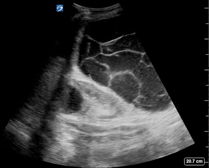
Image 1: Right upper quadrant FAST view; There is a complex cystic structure with internal loculations. There is some ascites and an incompletely visualized cystic structure involving the upper pole of the right kidney.
Image 2: Right upper quadrant, coronal view; Again there is a large complex cystic structure with numerous internal loculations. There is fine dependent, layering echogenic debris that has a relatively homogeneous ground glass appearance.
Image 3: Right abdomen, transverse view. The gravity dependent hyperechoic debris is again seen.
Fine layering echogenic debris is generally blood or pus. In this case with rapid onset, no features of infection and in an anticoagulated patient blood is almost certainly the cause. The structure is rounded and appears contained rather than free. Retroperitoneal bleeding or a contained intraperitoneal bleed are likely.
Image 4: This is a CT scan of the same patient.
CLINICAL CORRELATION
Retroperitoneal haemorrhage in anticoagulated patient.
Ultrasound has a poor sensitivity for diagnosing retroperitoneal haemorrhage, but in this patient the huge retroperitoneal collection is easily visualized.
CT confirmed this was a large retroperitoneal haemorrhage displacing the right kidney cranially, with ongoing active bleeding.
There was also a large amount of ascites.
[cite]
TOP 100 ULTRASOUND CASES


