Ultrasound Case 062
Presentation
A 30 year old woman presents with severe epigastric pain. The non-steroidal anti-inflammatory drugs she had taken hadn’t helped.
View 2
Describe and interpret these scans
IMAGE INTERPRETATION
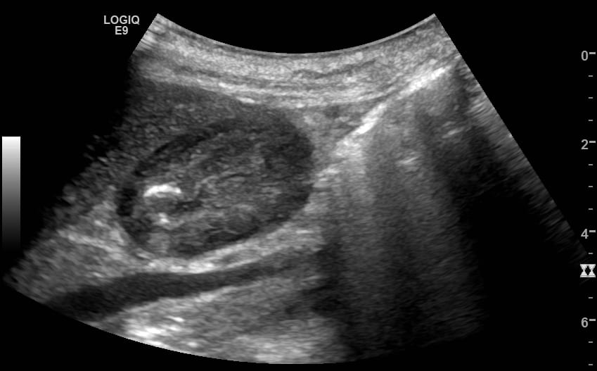
Image 1: Short axis view of the gastric antrum moving from pylorus toward body of the stomach.
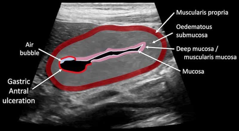
The layers of the gastric wall are clearly seen, and there is marked thickening particularly of the submucosa. There is a punched out lesion in which a small gas bubble sits seen at 9 o’clock when viewing the antrum in cross section. This is an antral peptic ulcer with marked associated submucosal oedema.
Image 2: Long axis view of the thick wall gastric antrum. The aorta, IVC, SMA and SMV and vertebral body of L1 are seen posterior to the antrum.
Image 3: Short axis view of the gastric antrum with high resolution linear transducer.
Image 4 & 5: Curvilinear still images of Image 1 & 2.
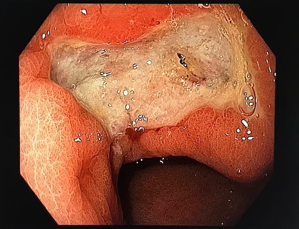
Image 6: Endocopic image demonstrating 20mm punched out gastric antral ulcer with surrounding oedematous antral wall.
CLINICAL CORRELATION
Helicobacter associated gastritis and peptic ulcer with associated submucosal oedema
Whilst not the investigation of choice ultrasound can sometimes show features of peptic ulcer disease. Gastric wall oedema and an ulcer crater are the characteristic findings and were seen in this case.
Optimal ultrasound imaging of the gastric antrum and duodenum is done by getting the patient to ingest water, then turn to the right lateral decubitus position. Antral wall thickness of ≤ 4mm is considered normal.
Diagnosis should be confirmed with endoscopy and consideration given to biopsy as a thickened gastric wall is non-specific and may be associated with peptic ulcer disease, gastritis – infectious, eosinophilic or inflammatory, malignancy particularly lymphoma, or Ménétrier disease (idiopathic hypertrophic gastropathy).
[cite]
TOP 100 ULTRASOUND CASES
An Emergency physician based in Perth, Western Australia. Professionally my passion lies in integrating advanced diagnostic and procedural ultrasound into clinical assessment and management of the undifferentiated patient. Sharing hard fought knowledge with innovative educational techniques to ensure knowledge translation and dissemination is my goal. Family, wild coastlines, native forests, and tinkering in the shed fills the rest of my contented time. | SonoCPD | Ultrasound library | Top 100 | @thesonocave |


