![]()
A Curtain Descends
aka Ophthalmology Befuddler 008
A 50 year-old man presents with loss of vision. He describes a curtain coming down across his vision. It was preceded by ‘flashes and floaters’.

Questions
Q1. What is the diagnosis?
Answer and interpretation
Retinal detachment
This is the separation of the sensory retina from the underlying pigmented retinal epithelium.
Findings:
- ultrasound — The detached retina is visible as a free floating echogenic membrane separated from the globe posteriorly. It moves with eye movement and is attached at the optic disc.
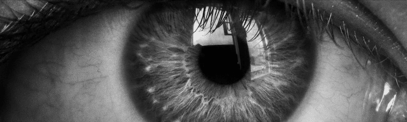
- ophthalmoscopy — The detached retina appears corrugated and partially opaque. On funduscopy the detached portion will appear out of focus.
Q2. What are the 3 types of mechanisms that can cause this condition?
Answer and interpretation
There are 3 types of retinal detachment:
- rhegmatogenous — the detached retina is elevated by underlying fluid that collects from the vitrous through a tear in the retina. This is the most common mechanism. It may be related to trauma, but is more common in men, those over age 45 years and those with myopia.
- exudative — fluid collects from retinal vessels. The causes may be neoplastic, inflammatory, congenital, or vascular in nature and include hypertension, prececlampsia, central retinal venous occlusion (CRVO), glomerulonephritis, papilledema, vasculitis, and choroidal tumours.
- tractional — the retina is pulled up by fibrocellular bands. This occurs in conditions such as proliferative diabetic retinopathy, sickle cell disease, retinopathy of prematurity(ROP), previous vitreous hemorrhage, trauma, and toxocariasis.
Q3. What are the features on history and examination?
Answer and interpretation
History:
- painless loss of vision (central, peripheral or both)
- Recent history of increased numbers of flashes (due to traction on the retina) and floaters (due to hemorrhage and debris in the vitreous).
- presence of a dark shadow or curtain moving over the visual field of the affected eye.
Examination:
- Visual acuity — reduced if the macula is involved.
- Red reflex — abnormal; a mobile detached retina may be visible.
- Visual fields — reduced.
- Pupils — a mild relative afferent pupillary defect (RAPD) may be present depending the size of the retinal detachment.
- Ophthalmoscopy — The detached retina appears corrugated and partially opaque. On funduscopy the detached portion will appear out of focus.
Other features that may be seen include: anterior vitreous pigmented cells, vitreous hemorrhage, and posterior vitreous detachment.
The slit lamp and ultrasound findings are shown in a short but enlightening video at RootAtlas
a
Q4. Describe investigation and management.
Answer and interpretation
Direct funduscopy in the emergency department cannot rule out retinal detachment — ultrasound is a useful investigation for diagnosing retinal detachment in the ED.
- Urgent ophthalmologist opinion.
- minimise activity —- bed rest with toilet privileges.
- Treatment of underlying cause (especially if exudative).
- Surgical options include laser photocoagulation, cryotherapy, pneumatic retinopexy, vitrectomy, and scleral buckle.
- Close follow up is required.
Q5. What is a retinal break?
Answer and interpretation
A retinal break is a tear in the retinal membranes and may or may not lead to retinal detachment.
Q6. What is retinoschisis?
Answer and Interpretation
Retinoschisis should be considered in the differential of retinal detachment. It refers to the splitting of the retina, which has X-linked juvenile and age-related degenerative forms.
It may be asymptomatic or lead to vision loss due to macular involvement and vitreous hemorrhage. It may be amenable to surgery.
References
- LITFL Top 100 – Case 38 Retinal detachment
- Ehlers JP, Shah CP, Fenton GL, Hoskins EN. The Wills Eye Manual: Office and Emergency Room Diagnosis and Treatment of Eye Disease Lippincott Williams & Wilkins
- NSW Statewide Opthalmology Service. Eye Emergency Manual — An illustrated Guide. [Free PDF]
OPHTHALMOLOGY BEFUDDLER
Chris is an Intensivist and ECMO specialist at The Alfred ICU, where he is Deputy Director (Education). He is a Clinical Adjunct Associate Professor at Monash University, the Lead for the Clinician Educator Incubator programme, and a CICM First Part Examiner.
He is an internationally recognised Clinician Educator with a passion for helping clinicians learn and for improving the clinical performance of individuals and collectives. He was one of the founders of the FOAM movement (Free Open-Access Medical education) has been recognised for his contributions to education with awards from ANZICS, ANZAHPE, and ACEM.
His one great achievement is being the father of three amazing children.
On Bluesky, he is @precordialthump.bsky.social and on the site that Elon has screwed up, he is @precordialthump.
| INTENSIVE | RAGE | Resuscitology | SMACC