![]()
Alexis Littré
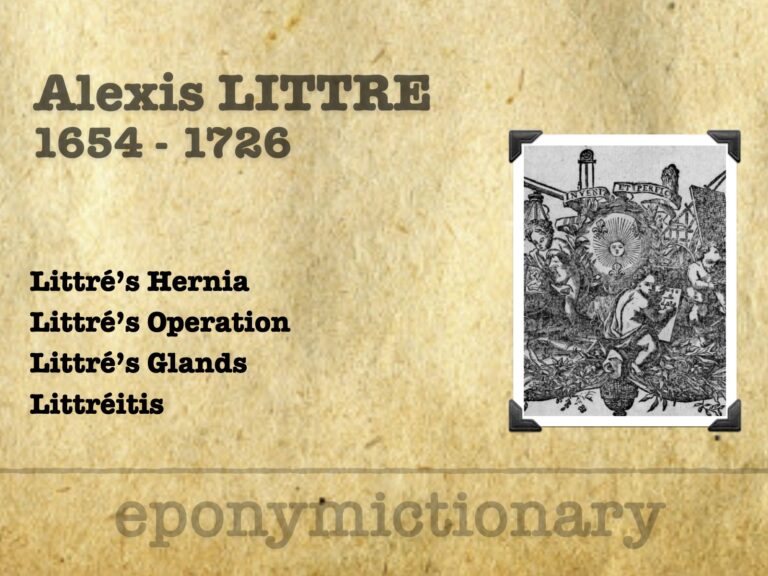
Alexis Littré (1654–1726), French anatomist; Littré’s hernia, glands, and operation; anatomical insights with lasting surgical impact

![]()
Alexis Littré (1654–1726), French anatomist; Littré’s hernia, glands, and operation; anatomical insights with lasting surgical impact
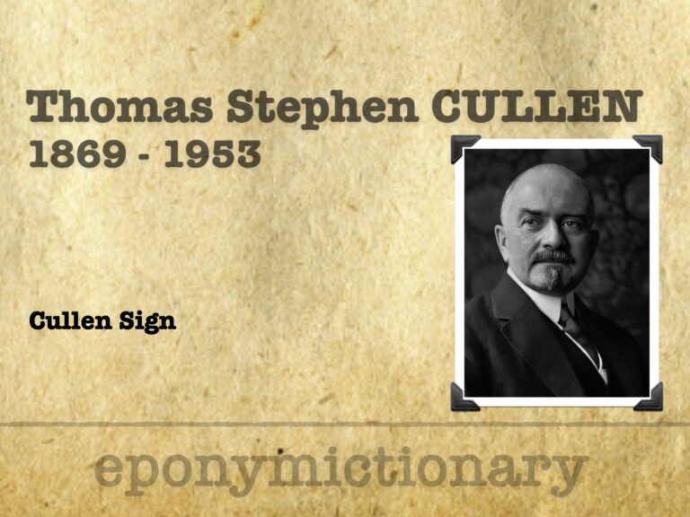
Thomas Stephen Cullen (1869 – 1953) was a Canadian gynecologist. Eponymously affiliated with Cullen sign (1918)
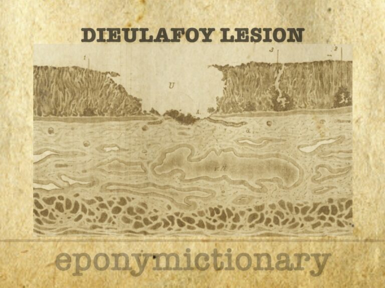
Dieulafoy’s lesion: minute gastric erosion over a large arteriole, causing massive GI bleeding. First defined as exulceratio simplex in 1898.

August Gottlieb Richter (1742–1812), German surgeon; Richter’s hernia, advanced cataract extraction, and elevated surgery into academia

Sir William Stokes (1839–1900), Irish surgeon and son of William Stokes, pioneered surgical techniques and served as RCSI professor and Queen Victoria’s surgeon
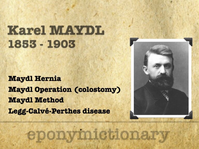
Karel Maydl (1853–1903), Czech surgeon, pioneer of colostomy, bladder exstrophy surgery, and Maydl’s hernia; early describer of Legg-Calvé-Perthes disease (LCPD)
John Henry Bryant (1867–1906) English physician. Eponym: Blue Scrotum Sign of Bryant associated with ruptured abdominal aortic anurysm (1903)

René-Jacques Croissant de Garengeot (1688–1759), Parisian surgeon, described appendix in femoral hernia, wrote on lacrimal surgery, and devised the tooth key
Charles Heber McBurney (1845 – 1913) was an American surgeon. Most famous for McBurney's point (1889) and McBurney's incision (1894) Medical Eponym.

Non-traumatic abdominal ecchymosis of the abdominal wall and flanks (Grey Turner, Cullen and Stabler); scrotum (Bryant) and upper thigh (Fox) as clues to potentially serious causes of abdominal pathology.

James Sherren (1872-1945) British General surgeon. Eponym: Sherren's triangle - area of hyperaesthesia associated with appendicitis
Adriaan van den Spiegel (1578–1625), Flemish anatomist; described Spigelian line, fascia, hernia, and liver lobe in his posthumous atlas.